U.S. flag

An official website of the United States government

NCBI Bookshelf. A service of the National Library of Medicine, National Institutes of Health.

Adam MP, Feldman J, Mirzaa GM, et al., editors. GeneReviews® [Internet]. Seattle (WA): University of Washington, Seattle; 1993-2024.

Cover of GeneReviews®

GeneReviews® [Internet].

Show details

Fibrous Dysplasia / McCune-Albright Syndrome

Synonym: FD/MAS

, MD, , MD, , PhD, , MD, and , MD.

Author Information and Affiliations

Initial Posting: ; Last Update: February 8, 2024.

Estimated reading time: 53 minutes

Summary

Clinical characteristics.

Fibrous dysplasia / McCune-Albright syndrome (FD/MAS), the result of an early embryonic postzygotic somatic activating pathogenic variant in GNAS (encoding the cAMP pathway-associated G protein Gαs [Gs alpha subunit]), is characterized by involvement of the skin, skeleton, and certain endocrine organs. However, because Gαs signaling is ubiquitous, additional tissues may be affected.

Hyperpigmented skin macules are common and are usually the first manifestation of the disease, apparent at or shortly after birth. Fibrous dysplasia (FD), which can involve any part and combination of the craniofacial, axial, and/or appendicular skeleton, can range from an isolated, asymptomatic monostotic lesion discovered incidentally to severe, disabling polyostotic disease involving practically the entire skeleton and leading to progressive scoliosis, facial deformity, and loss of mobility, vision, and/or hearing. Endocrinopathies include gonadotropin-independent precocious puberty resulting from recurrent ovarian cysts in girls and autonomous testosterone production in boys; testicular lesions with or without associated gonadotropin-independent precocious puberty; thyroid lesions with or without non-autoimmune hyperthyroidism; growth hormone excess; FGF23-mediated phosphate wasting with or without hypophosphatemia in association with fibrous dysplasia; and neonatal hypercortisolism.

Diagnosis/testing.

In most individuals, the diagnosis of FD/MAS is based on the finding of two or more typical clinical features. In individuals whose only clinical finding is monostotic FD, identification of a somatic activating pathogenic variant in GNAS by molecular genetic testing is required to establish the diagnosis. Variant detection depends on the level of mosaicism in the tissue and the sensitivity of the test method.

Management.

Treatment of manifestations: Management is most effectively accomplished by a multidisciplinary team of specialists. FD: management focuses on optimizing function and minimizing morbidity related to fractures and deformity (including scoliosis). Precocious puberty: treatment prevents bone age advancement and compromise of adult height. For girls, the aromatase inhibitor letrozole is used; for boys, treatment options are less well established. Thyroid disease: methimazole effectively manages hyperthyroidism; however, because hyperthyroidism is persistent, thyroidectomy is common. Growth hormone excess: medical therapy is the preferred first-line treatment; options include (alone or in combination) octreotide and the growth hormone receptor antagonist pegvisomant. Hypercortisolism: treatment varies by the presentation of neonatal Cushing syndrome.

Surveillance: FD/MAS: in infants, monitor for clinical signs of hypercortisolism; in all children, monitor for growth acceleration and other clinical signs of precocious puberty and/or growth hormone excess; in children age <5 years, monitor for thyroid function abnormalities; in those with thyroid abnormalities on ultrasound examination but normal thyroid function, perform periodic monitoring of thyroid function; in males, monitor for testicular lesions (physical examination and testicular ultrasound); in individuals on pegvisomant, monitor for hepatotoxicityl in those on homatostatin analogs, monitor for signs and symptoms of gallbladder disease; in females, monitor for breast cancer (earlier than is recommended for the general population). FD: periodic radiographs to monitor existing FD and development of new lesions; periodic serum phosphorus (for development of hypophosphatemia) and 25-hydroxyvitamin D levels. Craniofacial FD: yearly vision and hearing evaluations; periodic skull CT; routine serum IGF-1 levels through young adulthood. Spine FD: close monitoring for progressive scoliosis.

Agents/circumstances to avoid: Contact sports and other high-risk activities (when skeletal involvement is significant); prophylactic optic nerve decompression (in individuals with craniofacial FD); surgical removal of ovarian cysts; radiation therapy for treatment of FD; risk factors for malignancy (e.g., radiation exposure).

Genetic counseling.

FD/MAS is not inherited. No parent of a child with FD/MAS has been demonstrated to have any significant, distinctive manifestations of the disorder. The risk to sibs is expected to be the same as in the general population. There are no verified instances of vertical transmission of FD/MAS.

Diagnosis

Fibrous dysplasia / McCune-Albright syndrome (FD/MAS) is usually diagnosed based on characteristic clinical, radiographic, and laboratory manifestations, although formal diagnostic criteria have not been published.

Suggestive Findings

FD/MAS should be suspected in individuals with any of the following skin, skeletal, or endocrine features.

Skin. Individuals may have characteristic hyperpigmented skin macules. These have been referred to as café au lait macules; however, this does not accurately reflect their appearance on darker-skinned individuals.

  • Borders are jagged and irregular, often referred to as resembling the "coast of Maine" (in contrast to the smooth-bordered "coast of California" lesions seen in neurofibromatosis type 1).
  • Distribution shows an association with ("respecting") the midline of the body and following the developmental lines of Blaschko, which reflect patterns of embryonic cell migration (see Figure 1).
Figure 1.

Figure 1.

Hyperpigmented macules A. Skin lesions in a newborn demonstrating the characteristic association with the midline of the body, and distribution reflecting patterns of embryonic cell migration (developmental lines of Blaschko)

Skeletal. Fibrous dysplasia (FD), in which normal bone and bone marrow is replaced by fibro-osseous tissue, results in an increased risk of fractures, deformity, functional impairment, and pain.

  • FD can be monostotic (i.e., involvement of one bone) or polyostotic (i.e., involvement of >1 bone).
  • FD can involve any part and combination of the craniofacial, axial, and/or appendicular skeleton (see Figure 2).
  • The initial radiologic evaluation for FD should include a 99Tc-MDP (technetium-99 conjugated with methylene diphosphonate) or 18F-NaF (fluorine-18-labeled sodium fluoride) PET/CT bone scan. Areas of skeletal involvement identified on scintigraphy should be further evaluated with radiographs and head CT, depending on the location and extent of the disease (see Figure 3).
Figure 2.

Figure 2.

Fibrous dysplasia (FD) A. FD of the proximal femur demonstrating the typical "ground-glass" appearance with a coxa vara ("shepherd's crook") deformity

Figure 3.

Figure 3.

Suggested evaluations to determine if fibrous dysplasia (FD) is present and the extent of disease if FD is present 99Tc-MDP = technetium-99 conjugated with methylene diphosphonate; FD = fibrous dysplasia; H&P = history and physical exam; TRP = (more...)

Endocrine. Findings may include the following:

  • Gonadotropin-independent precocious puberty
  • Testicular lesions including Leydig and/or Sertoli cell hyperplasia with characteristic ultrasonographic features, with or without associated gonadotropin-independent precocious puberty (See Figure 4B.)
  • Thyroid lesions with characteristic ultrasonographic features, with or without non-autoimmune hyperthyroidism (See Figures 4C and 4D.)
  • Growth hormone excess
  • Fibroblast growth factor 23 (FGF23)-mediated phosphate wasting with or without hypophosphatemia
  • Neonatal hypercortisolism
Figure 4.

Figure 4.

Ultrasonography findings of ovarian, testicular, and thyroid disease A. Pelvic ultrasound in a girl age seven years, showing a complex unilateral ovarian cyst (defined by crosshatches). The uterus is prepubertal in size (arrow).

Establishing the Diagnosis

The clinical diagnosis of FD/MAS is established in individuals who have two or more typical clinical features of FD/MAS. In individuals whose only clinical finding is monostotic FD, identification of a somatic activating GNAS pathogenic variant is required to confirm the diagnosis (see Table 1).

Molecular genetic testing approaches include targeted analysis of codons p.Arg201 and p.Gln227. Molecular testing of affected tissue has the highest clinical sensitivity in PCR-sequencing-based diagnostic methods:

  • ~80% in lesional tissue
  • ~20%-30% in peripheral blood leukocytes

Note: (1) Variant detection depends on the level of mosaicism in the tissue and the sensitivity of the technique. Detection frequency of a variant at p.Arg201 using standard PCR was highest in endocrine organs and lowest in affected skin specimens [Lumbroso et al 2004]. The ability to detect mosaicism affects the detection rate of the assay (see Table 1 and Table 6). (2) Targeted analysis may be performed by sequencing GNAS exons 8 and 9. GNAS variants other than those previously reported to be associated with FD/MAS would likely be interpreted as variants of uncertain significance. (3) The cAMP pathway-associated G protein Gαs (Gs alpha subunit) is expressed in nearly all tissues from both maternal and paternal GNAS alleles. However, GNAS is a complex locus where alternative transcripts and additional phenotypes may result from GNAS imprinting (see Genetically Related Disorders and Molecular Genetics).

Table 1.

Molecular Genetic Testing Used in Fibrous Dysplasia / McCune-Albright Syndrome

Gene 1MethodVariants DetectedProportion of Probands with a Pathogenic Variant 2 Detectable by Method
GNAS Targeted analysis of exons 8 & 9 in affected tissue 3,4p.Arg201His, p.Arg201Cys 5, 6
  • 8%-90% 7
  • 75%-100% 8
p.Gln227Leu 6<5% 5
1.
2.

See Molecular Genetics for information on variants detected in this gene.

3.

Targeted analysis may be performed by sequence analysis. Sequence analysis detects variants that are benign, likely benign, of uncertain significance, likely pathogenic, or pathogenic. For issues to consider in interpretation of sequence analysis results, click here.

4.

Testing tissue from a lesion biopsy has a higher clinical yield than testing a blood sample. The detection rate for a blood sample is ~20%-30% [Lumbroso et al 2004, Kalfa et al 2006].

5.

Somatic GNAS missense variants in individuals with FD/MAS are known to occur at only one of two amino acid residues: p.Arg201 (>95% of pathogenic variants) [Lumbroso et al 2004] or p.Gln227 (<5%) [Idowu et al 2007].

6.

Rarely, other amino acid substitutions at p.Arg201 and at p.Gln227 have been detected (see Molecular Genetics).

7.

Variant detection depends on the level of mosaicism in the tissue and the sensitivity of the technique. Variant detection at p.Arg201 using standard PCR was highest in endocrine organs and lowest in affected skin specimens [Lumbroso et al 2004].

8.

When modified primers (peptide nucleic acid) [Bianco et al 2000] and next-generation sequencing [Narumi et al 2013] technologies are combined [Narumi et al 2013], a p.Arg201 variant can be detected in virtually all affected tissues and in leukocytes in up to 75% of individuals.

Clinical Characteristics

Clinical Description

Fibrous dysplasia / McCune-Albright syndrome (FD/MAS) results from mosaic somatic activating pathogenic variants in GNAS, which encodes the cAMP pathway-associated G protein Gαs (Gs alpha subunit). Affected tissues can include those derived from ectoderm, mesoderm, and endoderm, and commonly include skin, skeleton, and certain endocrine organs. However, because Gαs signaling is present in virtually every tissue, additional sites may be affected.

The phenotypic spectrum of FD/MAS ranges from asymptomatic incidental findings to neonatal lethality. There is a high degree of variability between individuals, both in the number of affected tissues and the degree to which they are affected. Disease manifestations depend on the time during embryogenesis that the somatic pathogenic variant occurred, the tissue involved, and the role of Gαs in the affected tissue. Pathogenic variants occurring early in development lead to widespread disease, while those occurring later in development lead to limited disease.

Hyperpigmented macules are common and are usually the first manifestation of FD/MAS, apparent at or shortly after birth. There is no correlation between the size of the skin lesions and the extent of disease, nor between the distribution of skin lesions and the location of fibrous dysplasia.

Fibrous dysplasia (FD) demonstrates a mosaic pattern: it can involve any part or combination of the craniofacial, axial, and/or appendicular skeleton. The bones most commonly involved are the skull base and proximal femurs [Kelly et al 2008]. While there is generally a central-to-peripheral gradient, any combination of involved bones is possible. FD is widely variable, from an asymptomatic monostotic lesion discovered incidentally to severe, disabling polyostotic disease involving practically the entire skeleton and leading to loss of vision, hearing, and/or mobility.

Individual bone lesions typically manifest during the first few years of life and expand during childhood. The vast majority of clinically significant bone lesions are detectable by age ten years, with few new and almost no clinically significant bone lesions appearing after age 15 years [Hart et al 2007]. In adulthood, FD lesions typically become less active, likely related to apoptosis of pathogenic variant-bearing cells [Kuznetsov et al 2008].

The clinical presentation and course of FD depends on the location and extent of the affected skeleton:

  • Appendicular skeleton. Children with FD in the appendicular skeleton typically present with a limp, pain, and/or pathologic fractures. Recurrent fractures and progressive deformity may lead to difficulties with ambulation and loss of mobility.
  • Craniofacial region. FD may present as a painless "lump" or facial asymmetry. Expansion of craniofacial lesions may lead to progressive facial deformity (see Figure 2B), and in rare instances (usually in association with growth hormone excess) loss of vision and/or hearing due to compromise of the optic nerves and/or external auditory canals [Cutler et al 2006, Boyce et al 2018].
  • Vertebrae. FD involving the vertebrae is common, and may lead to scoliosis, which in rare instances may be severe, progressive, and even lethal [Berglund et al 2018]. Untreated, progressive scoliosis is one of the few features of FD that can lead to early morbidity.

Bone pain is a common complication of FD. Although bone pain may present at any age, it is common for bone pain to be absent in childhood, occur in adolescence, and progress into adulthood [Kelly et al 2008].

Aneurysmal bone cysts are rapidly expanding fluid-filled lesions that form within preexisting areas of FD. Such lesions are best detected by MRI. Affected individuals experience acute onset of severe pain, rapidly expanding localized deformity, and rarely – when cysts compress the optic nerve – rapid loss of vision. Aneurysmal bone cysts thus carry a high risk of morbidity (see Management).

Malignant transformation of FD lesions is a rare complication. Many instances of malignant transformation were reported in association with previous radiation treatment [Ruggieri et al 1994]. Growth hormone excess may be a predisposing factor [Salenave et al 2014].

Radiographic appearance of FD varies according to location.

  • Radiographs of the appendicular skeleton show expansive lesions with endosteal scalloping, thinning of the cortex, and a "ground-glass" appearance (see Figure 2A).
  • FD in the craniofacial skeleton is typically expansile and appears sclerotic on radiographs, but demonstrates a typical "ground-glass" appearance on CT (see Figure 2C).
  • With aging, FD lesions in the appendicular skeleton tend to become sclerotic on radiographs, and craniofacial FD lesions develop a "cystic" appearance (see Figure 2D).

Endocrinopathies can include any of the following:

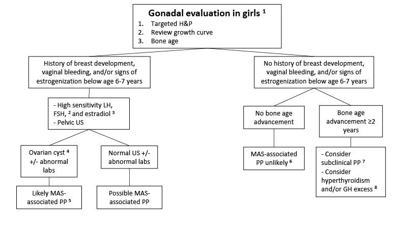
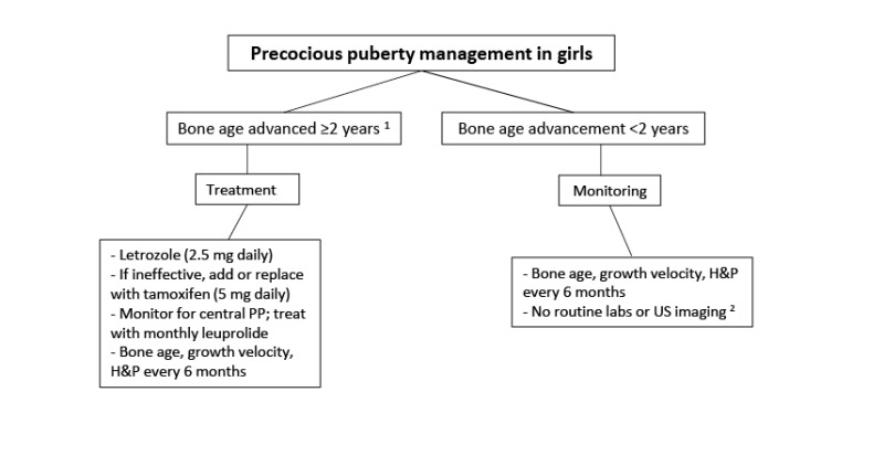
  • Precocious puberty is common in girls with FD/MAS (~85%) and is often the presenting feature. Recurrent ovarian cysts (see Figure 4A) lead to intermittent estrogen production resulting in breast development, growth acceleration, and vaginal bleeding; during the intervals between cyst formation, breast tissue typically regresses and estrogen levels fall to prepubertal levels. Ovarian cysts typically continue into adulthood, leading to irregular menses. This has the potential to interrupt ovulatory cycles, which may increase the time to conception in adult women. Ovarian torsion has been seen rarely in girls and women with large and persistent cysts [Clark et al 2000].
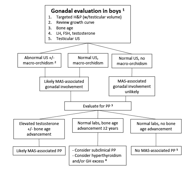
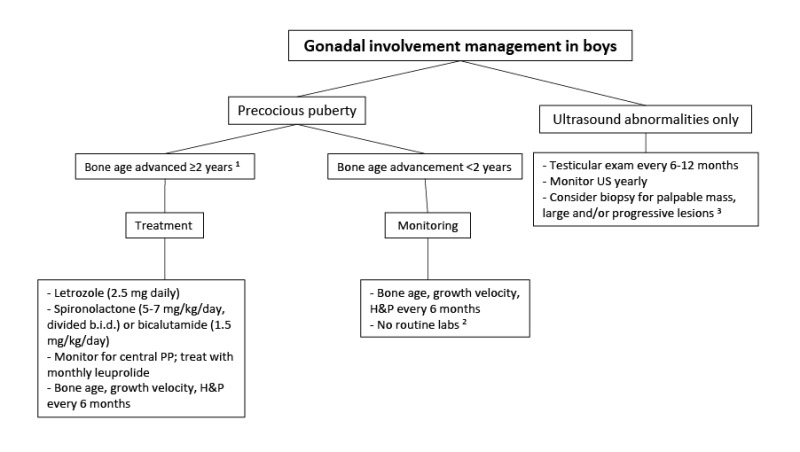
    Precocious puberty is less common in boys with FD/MAS (~10%-15%), and is due to autonomous testosterone production [Boyce et al 2012a], which leads to progressive pubertal development including growth acceleration, pubic and axillary hair, acne, and aggressive and/or inappropriately sexual behavior.
    In both girls and boys, prolonged autonomous sex steroid production typically leads to activation of the hypothalamic-pituitary axis and the development of central precocious puberty.
  • Fertility. The effects of autonomous sex steroid production on pituitary-gonadal function and fertility in adults are not well characterized. Women with FD/MAS may have recurrent ovarian cysts leading to irregular menses in adulthood [Lala et al 2007]. While many women in an NIH natural history study cohort have achieved successful pregnancies, it is possible that interruption of ovulatory cycles could decrease fertility and increase the time to conception [Boyce et al 2019].
  • Testicular abnormalities. Testicular abnormalities are seen in the majority of boys and men with FD/MAS (~85%), and typically manifest as unilateral or bilateral macro-orchidism [Boyce et al 2012a]. Ultrasound examination demonstrates discrete hyper- and hypoechoic lesions and microlithiasis, corresponding to areas of Leydig and/or Sertoli cell hyperplasia (see Figure 4B).
    The potential for malignant transformation of testicular lesions is unknown, but appears to be low [Boyce et al 2012a].
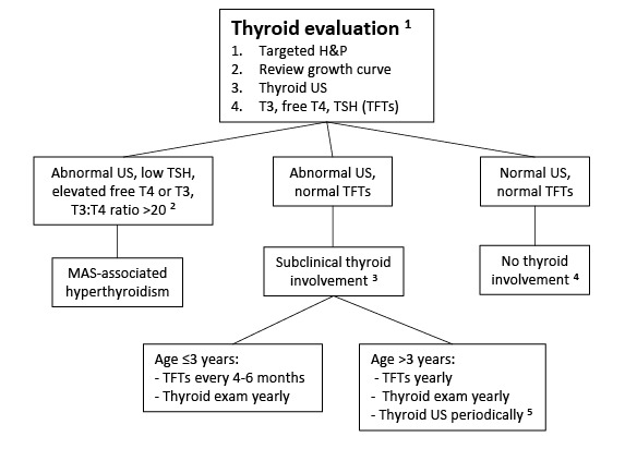
  • Thyroid disease. Thyroid involvement in FD/MAS is common. Approximately half of individuals with FD/MAS have ultrasound findings consistent with thyroid involvement, including mixed cystic and solid lesions interspersed with areas of normal-appearing tissue (see Figure 4C and 4D) [Celi et al 2008, Tessaris et al 2012].
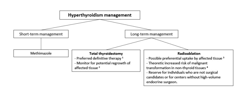
    Hyperthyroidism is present in 10%-30% of individuals with FD/MAS, and results from both increased hormone production and increased conversion of thyroxine (T4) to triiodothyronine (T3) [Celi et al 2008]. Hyperthyroidism is typically mild to moderate, but may be severe, and if undetected can lead to thyroid storm during anesthetic induction for surgery [Lawless et al 1992]. Uncontrolled hyperthyroidism may lead to bone age advancement, elevated bone turnover, and fractures.
    Malignant transformation of affected thyroid tissue has rarely been reported [Collins et al 2003, Hagelstein-Rotman et al 2021].
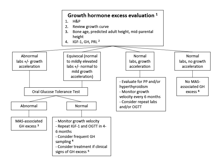
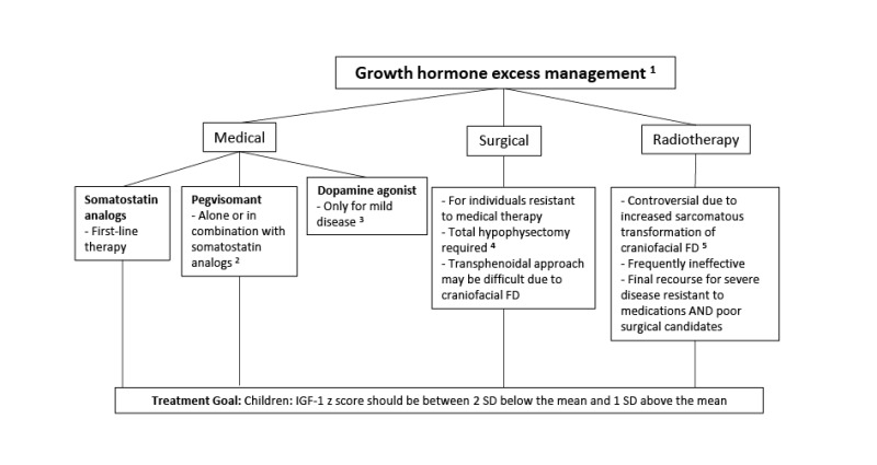
  • Growth hormone excess. Approximately 15%-20% of individuals with FD/MAS have GNAS pathogenic variants in the anterior pituitary that can lead to autonomous growth hormone production; approximately 80% of affected individuals with autonomous growth hormone production also have hyperprolactinemia [Salenave et al 2014].
    Affected individuals typically present with linear growth acceleration, and may develop features of acromegaly. Clinically, growth hormone excess must be distinguished from precocious puberty and hyperthyroidism, which also present with growth acceleration.
    Untreated growth hormone excess is associated with expansion of craniofacial FD, leading to macrocephaly and increased risk of vision and hearing loss [Boyce et al 2013, Boyce et al 2018] (see Figure 2B).
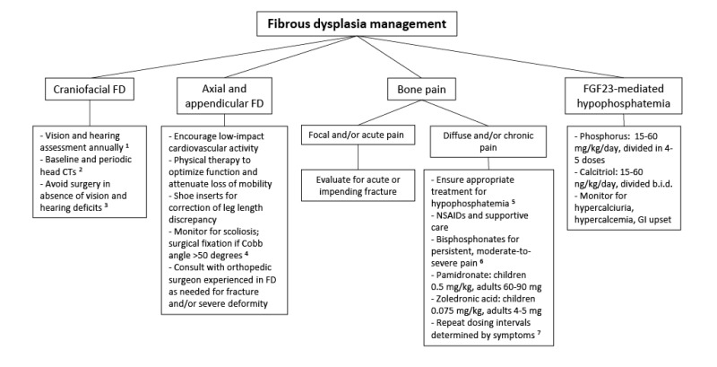
  • Fibroblast growth factor 23 (FGF23)-mediated phosphate wasting. In the majority of individuals with FD, increased production of the phosphaturic hormone FGF23 in FD tissue results in a renal tubulopathy with some degree of phosphate wasting [Collins et al 2001]. However, frank hypophosphatemia in persons with FD is infrequent, in part due to alterations in FGF23 processing that takes place in FD tissue and results in increased cleavage of FGF23 to its inactive fragments [Bhattacharyya et al 2012]. The degree of FGF23 overproduction in FD correlates with disease severity and skeletal burden; thus, frank hypophosphatemia is only seen in individuals with a substantial FD burden [Riminucci et al 2003].
    In contrast to most other features of FD/MAS, hypophosphatemia may wax and wane over the course of a person's lifetime and become more severe during periods of rapid skeletal growth. Hypophosphatemia may resolve as persons with FD become older, likely reflecting the intrinsic changes in FD that occur with age [Kuznetsov et al 2008].
    Affected individuals with frank hypophosphatemia may develop rickets/osteomalacia, increased fractures, skeletal deformities, and bone pain [Berglund et al 2018, Pan et al 2018, Geels et al 2022].
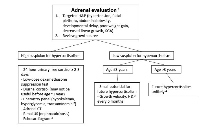
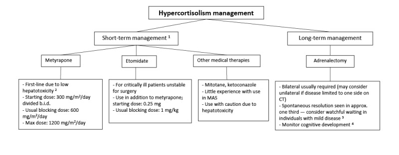
  • Hypercortisolism. Infants with FD/MAS may rarely present with Cushing syndrome due to excess cortisol production from the fetal adrenal gland [Brown et al 2010, Carney et al 2011]. Clinical symptoms typically develop in the neonatal period and may be severe, leading to critical illness and death. Spontaneous regression has been reported in approximately half of survivors, presumably related to fetal adrenal involution.

Liver manifestations

Gastrointestinal manifestations

  • Gastroesophageal reflux manifests in childhood and may be severe.
  • Upper gastrointestinal polyps have been described as a common finding in individuals with FD/MAS [Wood et al 2017].

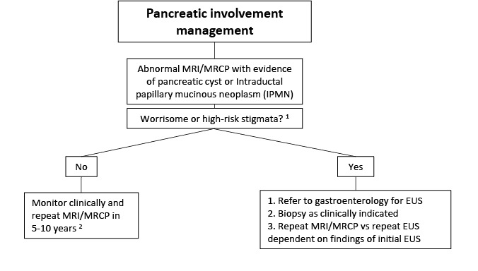
Pancreatic manifestations. Approximately 15% of individuals with FD/MAS have pancreatic complications:

  • Pancreatitis
  • Intraductal papillary mucinous neoplasms (IPMN) have been reported, which may present with variable grades of dysplasia [Gaujoux et al 2014, Wood et al 2017]. An individual with pancreatic carcinoma derived from an intestinal subtype of IPMN has been described [Parvanescu et al 2014].

Myxomas. Intramuscular myxomas are benign, usually asymptomatic, and often found incidentally. There are case reports positing a potential association between myxomas and increased risk of malignant transformation of FD [Biazzo et al 2017]; however, this has not been reported in larger series [Majoor et al 2019b, Hagelstein-Rotman et al 2021] and may be an artifact of publication bias.

Hematologic manifestations. Bone and bone marrow are, to varying degrees, replaced by fibro-osseous tissue typically devoid of hematopoietic marrow. There have been reports of bone marrow failure with pancytopenia and extramedullary hematopoiesis requiring splenectomy in individuals with FD/MAS [Mahdi et al 2017, Robinson et al 2018a].

Breast cancer. The risk for breast cancer in women with FD/MAS may be increased and can occur at a younger age compared to the general population. However, pathogenic activating GNAS variants were identified in only half of the breast tumors from women with FD/MAS studied [Majoor et al 2018a].

Health-related quality of life. Several series have shown impaired physical functioning in individuals with FD/MAS, strongly correlated with disease severity. There is conflicting evidence pertaining to the impact of the condition on psychosocial functioning, and this requires further invesigation [Kelly et al 2005, Majoor et al 2018b, Konradi 2021, Meier et al 2022].

Genotype-Phenotype Correlations

To date, only activating GNAS somatic pathogenic variants at residues p.Arg201 and p.Gln227 have been identified in individuals with FD/MAS. There are no known genotype-phenotype correlations [Zhadina et al 2021]. Disease severity is likely correlated with the degree of mosaicism and the tissues that are affected.

Nomenclature

The association of intramuscular myxomas with FD/MAS has been termed "Mazabraud syndrome." However, myxomas are more accurately categorized as an established feature of McCune-Albright syndrome (MAS).

Prevalence

FD/MAS is rare. While reliable data of prevalence are not available, estimates range between 1:100,000 and 1:1,000,000.

In contrast, FD (particularly the monostotic form) is not rare, and has been estimated to account for as much as 7% of all benign bone tumors.

Differential Diagnosis

Table 3.

Genes of Interest in the Differential Diagnosis of Fibrous Dysplasia / McCune-Albright Syndrome

Gene(s)DisorderMOIFeatures of Disorder
Overlapping w/FD/MASDistinguishing from FD/MAS
HRAS
NRAS
Cutaneous-skeletal hypophosphatemia syndrome (CSHS) 1Not inherited 2
  • FGF23-mediated hypophosphatemia
  • Hyperpigmented macules that follow developmental lines of Blaschko
  • Skeletal features (e.g., skeletal deformities, dysplastic bone lesions, scoliosis, craniofacial involvement ranging from calvarial thinning & maxillary hypoplasia to severe osteolysis w/large calvarial defects)
  • Epidermal & congenital melanocytic nevi
  • Neurologic abnormalities
  • Endocrinopathies are not a common feature, but central precocious puberty, thyroid nodules, & pheochromocytoma have been reported.
  • Ophthalmologic disorders (colobomas, limbal dermoids, strabismus, corneal opacities)
NF1 Neurofibromatosis 1 (NF1)AD
  • ≥6 café au lait macules that are generally smooth bordered ("coast of California") as opposed to the irregularly bordered ("coast of Maine") lesions seen in FD/MAS
  • Skeletal features (e.g., kyphoscoliosis, sphenoid dysplasia, cortical thinning of long bones, & bowing & dysplasia, particularly of the tibia, which may result in pseudarthroses)
  • Tumors of the nervous system (e.g., neurofibromas & optic gliomas)
  • Pigmented iris hamartomas
  • Axillary freckling
SH3BP2 3 Cherubism ADFibro-osseous skeletal lesions (See Table 4.)
  • Symmetric fibro-osseous lesions are generally limited to the maxilla & mandible.
  • No extraskeletal manifestations

AD = autosomal dominant; FD/MAS = fibrous dysplasia / McCune-Albright syndrome; MOI = mode of inheritance

1.
2.

CSHS is a mosaic disorder resulting from postzygotic somatic activating pathogenic variants in HRAS or NRAS [Lim et al 2014].

3.

In approximately 80% of affected individuals, cherubism arises from a heterozygous pathogenic variant in SH3BP2.

Fibro-osseous skeletal lesions may have radiologic and/or histologic features similar to fibrous dysplasia. These lesions are typically solitary, are not associated with extraskeletal features, and do not contain pathogenic variants in GNAS.

Table 4.

Fibro-osseous Skeletal Lesions in the Differential Diagnosis of Fibrous Dysplasia / McCune-Albright Syndrome

Skeletal LesionFeatures
Giant cell tumors of bone
  • Acquired lesions w/histopathologic features similar to FD, incl proliferation of bone marrow stromal cells & the presence of multiple multinucleated giant cells
  • Typically benign, but may result in localized bone destruction & (rarely) metastases
Ossifying fibromas
  • Benign lesions typically affecting the mandible & maxillae & presenting w/local expansion of a firm, painless mass
  • Ossifying fibromas are generally more aggressive than craniofacial FD lesions & are treated w/surgical excision.
Osteofibrous dysplasia
  • Typically occur in children age <10 yrs & most commonly affect the anterior tibia
  • Children present w/painless localized swelling &, in rare instances, w/fracture or progressive deformity.
  • Radiographs show a well-circumscribed radiolucent lesion w/characteristic sclerotic rim along the intracortical surface.
Cherubism
  • Radiographic manifestations typically incl bilateral, multilocular, radiolucent areas w/in the mandible, usually located at the angles & rami. The coronoid processes are commonly involved, whereas the condyles are rarely affected.
  • Histologic manifestations of lesions in the mandible &/or maxilla: non-neoplastic fibrotic lesions that contain numerous multinuclear giant cells & occasionally cysts. Increase in osteoid & newly formed bone matrix is observed in the periphery.

FD = fibrous dysplasia

Management

Evaluations Following Initial Diagnosis

After the initial diagnosis, all individuals with fibrous dysplasia / McCune-Albright syndrome (FD/MAS) should be evaluated to determine the extent of disease. The presence of any features of FD/MAS should prompt more detailed clinical evaluation for additional manifestations. The authors recommend the following studies, if they have not already been completed.

Skeleton

  • Total body bone scintigraphy to identify and determine the extent of fibrous dysplasia (FD). The majority of clinically significant skeletal lesions are apparent on bone scan by age five years.
  • Imaging of identified areas of FD with radiographs (axial and appendicular FD) and/or CT (craniofacial FD) to more clearly evaluate the extent and anatomy of the lesions
  • Baseline ophthalmologic, otolaryngologic, and audiologic evaluations in persons with craniofacial FD
  • Skeletal evaluation (See Figure 3.)

Endocrine. A thorough history, physical examination, and review of a growth chart (if available) are recommended to evaluate for clinical signs of endocrinopathies.

  • Biochemical screening for hyperthyroidism, growth hormone excess (insulin-like growth factor 1 [IGF-1] level), and fibroblast growth factor 23 (FGF23)-mediated hypophosphatemia (See Figure 3 and Figure 5.)
  • In individuals with clinical signs or a previous history of precocious puberty: biochemical screening, pelvic ultrasound examination (in females), and bone age examination (See Figure 6 and Figure 7.)
  • Ultrasound examination of the thyroid gland and testes (in all males) to evaluate for subclinical disease (See Figure 7 and Figure 8.)
  • Testing for hypercortisolism in infants with clinical evidence of Cushing syndrome (hypertension, facial plethora, abdominal obesity, developmental delay, poor weight gain, decreased linear growth, small size for gestational age) (See Figure 9.)

Less common manifestations. Consideration should be given to the less common manifestations cited in Clinical Description with appropriate clinical evaluations and imaging/biochemical studies performed as indicated (see Figure 10 for gastrointestinal evaluation).

Figure 5.

Figure 5.

Recommended evaluations for growth hormone excess in individuals with fibrous dysplasia / McCune-Albright syndrome

GH = growth hormone; H&P = history and physical examination; IGF-1 = insulin-like growth factor 1; MAS = McCune-Albright syndrome; OGTT = oral glucose tolerance test; PP = precocious puberty; PRL = prolactin

1. To be performed at initial presentation in all individuals with MAS, regardless of clinical symptoms.

2. The majority of individuals with MAS-associated GH excess will have increased prolactin secretion [Salenave et al 2014].

3. Practitioners may consider pituitary MRI in individuals suspected of having MAS-associated GH excess; however, findings may be nonspecific and rarely change management [Salenave et al 2014].

4. There are a variety of techniques for frequent GH sampling. Collecting GH samples every 20 minutes for 12 hours from 8 PM to 8 AM, with a lack of nadir below 1.0 ng/mL, is considered consistent with GH excess.

5. In those with craniofacial FD it is prudent to have a low threshold for initiating treatment, as uncontrolled GH excess is associated with increased craniofacial morbidity [Boyce et al 2012b].

6. MAS-associated GH excess may rarely present as late as young adulthood; therefore, ongoing monitoring with periodic IGF-1 levels is prudent in those with significant craniofacial FD.

Figure 6.

Figure 6.

Recommended evaluations for gonadal abnormalities in females with fibrous dysplasia / McCune-Albright syndrome

FSH = follicle-stimulating hormone; GH = growth hormone; H&P = history and physical examination; LH = luteinizing hormone; MAS = McCune-Albright syndrome; PP = precocious puberty; US = ultrasound

1. To be performed at initial presentation in all girls with MAS, regardless of clinical symptoms.

2. Gonadotropins should be suppressed in those with precocious puberty, unless autonomous estrogen production has induced central precocious puberty [Collins et al 2012].

3. Estrogen production in MAS-associated precocious puberty is intermittent, and undetectable levels do not eliminate the possibility of disease.

4. Ovarian cysts are suggestive of MAS-associated precocious puberty; however, absence of cysts does not eliminate the possibility of disease [Authors, personal observation].

5. In isolated peripheral precocious puberty, the differential diagnosis includes estrogen-producing tumor. Evaluation for additional features of MAS may establish the diagnosis.

6. Unlike other features of MAS, autonomous ovarian activity may present at any time during infancy or childhood. Girls should continue to be monitored clinically for signs of peripheral precocious puberty; however, routine laboratory testing and imaging is not recommended.

7. Affected females may rarely present with intermittent ovarian activity with only subtle signs of estrogenization (mild intermittent breast development without vaginal bleeding).

8. Hyperthyroidism and GH excess may present with an advanced bone age compared to chronologic age.

Figure 7.

Figure 7.

Recommended evaluations for gonadal abnormalities in males with fibrous dysplasia / McCune-Albright syndrome

FSH = follicle-stimulating hormone; GH = growth hormone; H&P = history and physical examination; LH = luteinizing hormone; MAS = McCune-Albright syndrome; PP = precocious puberty; US = ultrasound

1. Performed at initial presentation in all boys with MAS, regardless of clinical symptoms.

2. Typical MAS-associated macro-orchidism presents with uniform, unilateral, or bilateral testicular enlargement without discrete palpable masses.

3. Precocious puberty is less likely to occur in males who do not have evidence of testicular involvement on ultrasound. The presence of macro-orchidism is typically associated with US abnormalities.

4. Hyperthyroidism and GH excess may present with an advanced bone age compared to chronologic age.

5. Autonomous testicular activity may present at any time during childhood. Boys should continue to be monitored clinically for signs of peripheral precocious puberty; however, routine laboratory testing and imaging is not recommended [Boyce et al 2012a].

Figure 8.

Figure 8.

Recommended evaluations for thyroid abnormalities in individuals with fibrous dysplasia / McCune-Albright syndrome

H&P = history and physical examination; MAS = McCune-Albright syndrome; T3 = triiodothyronine; T4 = thyroxine; TFTs = thyroid function tests; TSH = thyroid-stimulating hormone; US = ultrasound

1. To be performed at initial presentation in all individuals with MAS, regardless of clinical symptoms.

2. An elevated T3:T4 ratio is suggestive of autonomous T3 production in individuals with MAS [Celi et al 2008].

3. A small percentage of affected individuals with radiologic thyroid abnormalities and normal TFTs will develop hyperthyroidism at some point during childhood.

4. The absence of biochemical or radiologic thyroid abnormalities after age five years likely eliminates the possibility of MAS-associated thyroid disease, and no further routine monitoring is required.

5. MAS-associated thyroid disease is correlated with a slightly increased risk of thyroid cancer (see Surveillance). Those with radiologic thyroid abnormalities should be monitored with yearly physical examination and thyroid US every 2-5 years.

Figure 9.

Figure 9.

Recommended evaluations for adrenal gland dysfunction in individuals with fibrous dysplasia / McCune-Albright syndrome

CT = computerized tomography; H&P = history and physical examination; SGA = small for gestational age; US = ultrasound

1. To be performed at initial presentation in all individuals with MAS, regardless of clinical symptoms.

2. Liver disease is highly correlated with MAS-associated hypercortisolism.

3. Prognosis of hypercortisolism is negatively correlated with the presence of comorbid heart disease [Brown et al 2010]. Since hypercortisolism may lead to heart disease, the presence of hypercortisolism makes the prognosis for heart disease worse.

4. Hypercortisolism in MAS results from autonomous activity of the adrenal fetal zone, which involutes rapidly after birth and is typically gone by age one year [Carney et al 2011]. MAS-associated hypercortisolism is unlikely after age one year, and the risk is effectively gone after age three years [Brown et al 2010].

Figure 10.

Figure 10.

Recommended evaluations for gastrointestinal issues in individuals with fibrous dysplasia / McCune-Albright syndrome

GERD = gastroesophageal reflux disease; GI = gastrointestinal; H&P = history and physical examination; MAS = McCune-Albright syndrome; MRCP = magnetic resonance cholangiopancreatography; MRI = magnetic resonance imaging

1. To be performed at initial presentation in all individuals with MAS, regardless of clinical symptoms.

2. Age is not based upon clinical evidence, but on age at which affected individuals may undergo MRI/MRCP without requiring sedation, and should be individualized based on clinical judgment.

3. Age of onset of pancreatic cyst development is not established; therefore, clinical monitoring for gastrointestinal symptoms in these affected individuals is indicated.

4. Affected individuals should continue to be monitored clinically for new signs of gastrointestinal/pancreatic involvement, including pancreatitis and diabetes [Gaujoux et al 2014, Parvanescu et al 2014, Wood et al 2017].

Treatment of Manifestations

Management is most effectively accomplished through the input of a multidisciplinary team of specialists, including an endocrinologist, orthopedic surgeon, physiatrist, ophthalmologist, audiologist, endocrine surgeon, craniofacial surgeon, and clinical geneticist. A consensus statement from the FD/MAS international consortium on best practice management guidelines was published in 2019 [Javaid et al 2019].

Fibrous Dysplasia

There are no established medical therapies capable of altering the disease course in FD. Current management is focused on optimizing function and minimizing morbidity related to fractures and deformity. The primary elements of management include the following (see also Figure 11):

  • Orthopedic surgery to repair fractures and to prevent and correct deformities. A surgeon experienced in FD should be consulted, as approaches previously considered standard (e.g., curettage, grafting, external fixation) are frequently ineffective [Stanton et al 2012, Leet et al 2016].
  • Diagnosis and treatment of scoliosis is of particular importance, as it may be rapidly progressive and in rare instances may lead to fatal respiratory compromise. For this reason, all individuals with spinal FD should be monitored closely by an orthopedic surgeon or physiatrist for possible progression. Surgical fusion has been shown to be effective at stabilizing the spine [Leet et al 2004b, Mancini et al 2009].
  • Aneurysmal bone cysts, best detected by MRI, are rapidly expanding fluid-filled lesions that form within preexisting areas of FD. Affected individuals experience acute onset of severe pain, rapidly expanding localized deformity, and rarely – when cysts compress the optic nerve – rapid loss of vision. Aneurysmal bone cysts thus carry a high risk of morbidity and should be evaluated urgently by a surgeon [Lee et al 2012, Manjila et al 2013].
  • Optical coherence tomography (OCT) can be used to more accurately assess for developing optic neuropathy in individuals with FD, as retinal nerve fiber layer thickness has been shown to be a better indicator of optic neuropathy than conventional CT assessment of optic canal stenosis and optic nerve stretch [Pan et al 2020].
  • Prophylactic optic nerve decompression to reduce the risk of vision loss can in fact increase the risk of vision loss and is thus contraindicated [Lee et al 2002, Cutler et al 2006, Amit et al 2011].
  • Physical therapy to optimize function and attenuate loss of mobility is appropriate. Affected individuals with lower-extremity FD in particular may benefit from therapies to address hip girdle weakness, range of motion, and leg length discrepancies [Paul et al 2014].
  • Intravenous bisphosphonates such as zoledronic acid and pamidronate are usually effective at relieving bone pain. Dosing should be based on symptoms, not on a fixed interval or bone turnover markers. The oral bisphosphonate alendronate has been shown to be ineffective for treatment of bone pain [Boyce et al 2014].
  • Denosumab, a human monoclonal antibody to receptor activator of nuclear kappa-B ligand (RANKL), has been used in several individuals with FD, with an apparent significant reduction in bone turnover markers and lesion activity, and has demonstrated improvement in osteogenic cell maturation and bone formation [de Castro et al 2023]. However, it has also been associated with clinically significant disturbances of mineral metabolism both while on treatment and after discontinuation, particularly in younger individuals and those with high FD burden [Boyce et al 2012b, de Castro et al 2023]. For this reason, denosumab should only be used in centers with extensive experience in the treatment of individuals with FD, ideally in the context of a clinical study.
  • Burosumab, a human monoclonal antibody to FGF23, has been used in an individual with FD who demonstrated sustained normalization of serum phosphate and improvement in alkaline phosphatase levels along with clinical improvement in bone pain [Gladding et al 2021]. Further study is needed to determine the clinical utility of burosumab in individuals with FD.
  • Malignancy should remain a consideration for individuals with acute or rapidly expanding FD lesions, or with atypical radiographic features such as compromise of the bony cortex with an associated soft tissue mass.
Figure 11.

Figure 11.

Recommended management for fibrous dysplasia in individuals with fibrous dysplasia / McCune-Albright syndrome

b.i.d. = twice daily; CT = computerized tomography; FD = fibrous dysplasia; GI = gastrointestinal; NSAIDs = nonsteroidal anti-inflammatory drugs

1. Affected individuals should be evaluated yearly by a neuro-ophthalmologist; less frequently once stability is demonstrated. Those with evidence of optic neuropathy should be referred to an experienced craniofacial surgical team.

2. Repeat head CT approximately every five years, potentially sooner in younger individuals, those with severe disease, or if vision or hearing deficits develop [Boyce et al 2017].

3. Optic nerve encasement is common and usually asymptomatic. Prophylactic optic nerve decompression in the absence of optic neuropathy is contraindicated [Lee et al 2002, Amit et al 2011].

4. Scoliosis may be progressive and potentially fatal in severe cases. All affected individuals with scoliosis should be followed regularly by an orthopedic surgeon [Leet et al 2004b].

5. Inadequately treated hypophosphatemia may significantly worsen bone pain and must be addressed before considering bisphosphonates [Leet et al 2004a, Paul et al 2014].

6. Bisphosphonates have not been shown to affect disease progression and use should be limited to treatment of FD-related bone pain [Hart et al 2007, Boyce et al 2014].

7. Doses should be repeated as needed when pain returns rather than on a set dosing schedule. In absence of significant decrease in bone pain, bisphosphonate treatment should be discontinued.

Endocrinopathies

Precocious puberty. Treatment of precocious puberty is important to prevent bone age advancement and compromise of adult height.

  • Females (see Figure 12). The aromatase inhibitor letrozole is an effective treatment for females [Feuillan et al 2007]. Studies have shown that letrozole treatment resulted in sustained beneficial effects on skeletal maturation, growth velocity, and predicted adult height [Estrada et al 2016, Wang & Yu 2018, Xu et al 2020]. Most individuals also have a decrease in the number of menstrual bleeding episodes while on treatment. Prophylactic surgical intervention for large and persistent ovarian cysts should be undertaken with extreme caution due to the known risk for cyst recurrence and the potential for decreased ovarian reserve in affected individuals.
  • Males (see Figure 13). Given the rarity of precocious puberty in boys, treatment options are less well established. One strategy includes the combination of an androgen receptor blocker (e.g., spironolactone or bicalutamide) and an inhibitor of sex steroid synthesis (e.g., letrozole) [Boyce et al 2012a].

Children of both sexes frequently enter central precocious puberty due to premature sex steroid exposure (see Clinical Description). This typically presents with reappearance of the signs of puberty in a child with previously well-controlled peripheral precocious puberty. Leuprolide therapy in combination with the above medications is an effective therapeutic strategy in most.

Figure 12.

Figure 12.

Recommended management for precocious puberty in girls with fibrous dysplasia / McCune-Albright syndrome

H&P = history and physical examination; PP = precocious puberty; US = ultrasound

1. The primary indication for treatment is to prevent impairment of adult height. Vaginal bleeding in the absence of bone age advancement does not typically warrant treatment. Exceptions may be made for very young children with frequent bleeding episodes deemed likely to lead to bone age advancement, or those who experience significant psychosocial distress related to pubertal episodes [Eugster et al 2003].

2. The primary end point for treatment efficacy is prevention of bone age advancement, which is assessed by growth velocity and bone age examination. Routine laboratory testing and US are unlikely to change management, and are not recommended.

Figure 13.

Figure 13.

Recommended management for gonadal involvement in boys with fibrous dysplasia / McCune-Albright syndrome

H&P = history and physical examination; b.i.d. = twice daily; PP = precocious puberty; US = ultrasound

1. The primary indication for treatment is to prevent impairment of adult height. Elevated testosterone levels in the absence of bone age advancement does not warrant treatment. Exceptions may be made for boys with testosterone-induced behavioral changes or progressive masculinization of the genitalia.

2. Routine laboratory testing is unlikely to change management and is not recommended.

3. Routine biopsy of affected testes is not recommended. Lesions should be followed with serial exam and US. Consider biopsy for lesions with atypical features such as a palpable mass, or for lesions that are large and/or progressive [Boyce et al 2012a].

Thyroid disease. Methimazole is effective for medical management of hyperthyroidism [Tessaris et al 2012] and is the first line of treatment. Propylthiouracil has been associated with an unacceptable risk of hepatotoxicity in children and therefore is no longer recommended [Ross et al 2016]. Because FD/MAS-associated hyperthyroidism is persistent, most affected individuals ultimately elect for definitive treatment. Thyroidectomy is the preferred definitive treatment in most affected individuals. Total gland resection is generally recommended due to the potential for thyroid tissue regrowth. Treatment by a highly experienced endocrine surgeon is critical to minimize complications and optimize outcomes. Affected individuals should be monitored postsurgically with yearly ultrasound examination to evaluate for tissue regrowth (see Figure 14.)

Radioablation is typically avoided due to potential preferential uptake by tissues bearing a somatic activating GNAS pathogenic variant, which may lead to increased risk of malignancy in the remaining unaffected gland. Additionally, GNAS pathogenic variants are associated with a slight increased risk of malignant transformation in both thyroid and non-thyroidal tissues; the risk is potentially enhanced by radiation exposure.

Figure 14.

Figure 14.

Recommended management for hyperthyroidism in individuals with fibrous dysplasia / McCune-Albright syndrome

1. Total thyroidectomy is preferred over subtotal as any remaining abnormal tissue has the potential to regrow, with recurrence of hyperthyroidism. Accordingly, radioactive iodine uptake scan will not alter management and is not part of routine preoperative care.

2. After thyroidectomy affected individuals should continue to be monitored with yearly physical exam and thyroid ultrasound.

3. Preferential uptake of radioactive iodine by diseased tissue may lead to a theoretic risk of thyroid cancer in the remaining unaffected tissue.

4. Both thyroid and non-thyroidal tissues with an activating pathogenic GNAS variant carry a slight increased risk of malignant transformation, which may be increased by radiation exposure [Collins et al 2003, Tessaris et al 2012].

Growth hormone (GH) excess. Medical therapy is the preferred first-line treatment. Options include (alone or in combination) somatostatin analogs and the GH receptor antagonist pegvisomant [Boyce et al 2013, Salenave et al 2014] (see Figure 15).

  • In growing children, the therapeutic goal is to maintain the insulin-like growth factor 1 (IGF-1) level in the middle of the normal range with an IGF-1 z score below zero.
  • In skeletally mature individuals, the goal is to decrease the IGF-1 level to as low as possible.

Medical therapy is typically continued indefinitely, as options for definitive treatment are associated with significant morbidity. Surgery may be technically difficult or precluded due to craniofacial FD. Additionally, given the diffuse pituitary infiltration of GH-producing cells, affected individuals treated surgically require total hypophysectomy with resulting total hypopituitarism [Vortmeyer et al 2012]. Radiation treatment may be effective in individuals with refractory disease, but has been associated with fatal malignant transformation of craniofacial FD [Hansen & Moffat 2003, Liu et al 2011].

The hyperprolactinemia that frequently accompanies GH excess is generally responsive to treatment with dopamine agonists, including cabergoline and bromocriptine. This class of drugs could also have an effect on GH excess treatment in those with modest elevations of GH and IGF-1 levels, with or without concomitant hyperprolactinemia [Katznelson et al 2014].

Figure 15.

Figure 15.

Recommended management for growth hormone excess in individuals with fibrous dysplasia / McCune-Albright syndrome

FD = fibrous dysplasia; GH = growth hormone; IGF-1 = insulin-like growth factor 1; SD = standard deviation

1. Hyperprolactinemia accompanies GH excess in approximately 80% of individuals with MAS. It usually only requires treatment if levels are very high and/or it is interfering with pubertal progression, menses, or sexual function [Salenave et al 2014].

2. The authors' practice is to add pegvisomant after reaching a maximal dose of somatostatin analogs.

3. Effective for treatment of modest elevations of GH and IGF-1 levels, with or without concomitant hyperprolactinemia.

4. Due to characteristic diffuse somatolactotroph hyperplasia of the pituitary, total hypophysectomy is required for successful surgical treatment [Vortmeyer et al 2012].

5. FD of the skull base is nearly universal in individuals with MAS-associated GH excess. There are reports of fatal skull base osteosarcomas arising after pituitary irradiation for treatment of MAS-associated GH excess [Liu et al 2011].

FGF23-mediated phosphate wasting. Treatment of frank hypophosphatemia is the same as in other disorders of FGF23 excess, and includes oral phosphorus and calcitriol. Important therapeutic end points include growth velocity and radiographic evidence of epiphyseal healing. Unlike other disorders of FGF23 excess, bone turnover markers in FD/MAS (e.g., alkaline phosphatase) may be constitutively elevated and are not a useful indicator of skeletal response to treatment.

Hypercortisolism. Treatment guidelines for hypercortisolism are difficult to establish given the rarity of neonatal Cushing syndrome. Additionally, affected individuals may be critically ill at presentation, which significantly affects treatment options.

  • Definitive treatment includes surgical removal of the diseased adrenal glands.
  • For medical treatment, metyrapone is frequently effective and is preferred over ketoconazole in children with liver abnormalities.

Spontaneous remission has been clearly documented in some affected individuals [Brown et al 2010]; however, it is not possible to identify prospectively which individuals will undergo remission. The decision to pursue or delay adrenalectomy must be made on an individual basis, taking into account the severity of illness, the ability of medications to control cortisol levels, and the potential effect of continued hypercortisolism on neurodevelopment (see Figure 16).

Figure 16.

Figure 16.

Recommended management for hypercortisolism in individuals with fibrous dysplasia / McCune-Albright syndrome

b.i.d. = twice daily; MAS = McCune-Albright syndrome; m2 = meters squared (referring to body surface area)

1. Affected individuals are often critically ill at presentation, which may impact treatment options.

2. Hepatotoxicity is an important consideration due to frequent comorbid liver disease [Brown et al 2010].

3. Spontaneous resolution may occur due to involution of the adrenal fetal zone, which is the source of hypercortisolism in MAS [Carney et al 2011].

4. Children with a current or remote history of MAS-associated hypercortisolism are at increased risk for neurodevelopmental delays, and should be considered for early interventional services [Brown et al 2010].

Pancreatic involvement. FD/MAS-associated intraductal papillary mucinous neoplasms (IPMNs) are common and tend to occur at a younger age than the general population. A study by Robinson et al [2018b] reported IPMNs in approximately 46% of their cohort, of which 40% had either worrisome or high-risk features. Pancreatic adenocarcinoma appears to be a rare development in this population [Parvanescu et al 2014]. Given the prevalence of IPMNs observed in this cohort, evaluation with MRI/MRCP should be considered for individuals with any concerning symptoms. Those with evidence of IPMNs should undergo surveillance, with recommendation to follow the guidelines for the evaluation of IPMNs in the general population [Tanaka et al 2017] (see Figure 17).

Figure 17.

Figure 17.

Recommended management for pancreatic involvement in individuals with fibrous dysplasia / McCune-Albright syndrome

EUS = endoscopic ultrasound; MRCP = magnetic resonance cholangiopancreatography; MRI = magnetic resonance imaging

1. Based on the 2012 International Consensus Guidelines for the management of intraductal papillary mucinous neoplasm (IPMNs) [Tanaka et al 2012]. Worrisome features include: pancreatitis; cyst ≥3 cm; thickened enhanced cystic walls; main pancreatic duct dilatation of 5-9 mm; non-enhanced mural nodules; and abrupt change in caliber of the main pancreatic duct with distal pancreatic atrophy, lymphadenopathy, an elevated serum level of carbohydrate antigen (CA) 19-9, and a rapid rate of cyst growth >5 mm within two years. High-risk stigmata on CT, MRI, or EUS include: obstructive jaundice in affected individuals with a cystic lesion in the pancreatic head; enhanced solid component (mural nodule) within the cyst (≥5 mm); and main pancreatic duct dilatation ≥10 mm.

2. The interval for repeat MRI/MRCP is not established [Gaujoux et al 2014, Wood et al 2017].

Surveillance

Due to the mosaic nature of FD/MAS, the clinical findings in affected individuals can vary significantly, with some individuals having involvement of only one organ system and others having more widespread involvement. Additionally, some features are age dependent and are either not likely to develop after a certain age or are more likely to affect older individuals. The following information on surveillance applies to individuals who have already been evaluated for signs and symptoms of the condition and in whom the extent of disease has been assessed; surveillance will need to be tailored to the individual's age and known affected organ systems (see Table 5).

Table 5.

Fibrous Dysplasia / McCune Albright Syndrome: Surveillance to Consider

System/ConcernEvaluationFrequency
Musculoskeletal 1 Monitoring for progression of scoliosis & other skeletal findings by orthopedic surgeon or physiatristRoutinely
CT of skullEvery 5 yrs or more frequently in younger persons, those w/severe disease, or if vision or hearing deficits develop
Radiographs to evaluate new or worsening symptoms & provide additional information about FD anatomy & bone qualityPeriodically
Endocrine Puberty (females) Eval for growth acceleration & other clinical signs of precocious puberty 2, 3At each visit
Bone age assessmentEvery 6 mos in those w/bone age advancement of ≥2 yrs
Puberty (males) Eval for growth acceleration & other clinical signs of precocious puberty 3, 4At each visit
Bone age assessmentEvery 6 mos in those w/bone age advancement of ≥2 yrs
Testicular physical examAt each visit
Testicular ultrasoundPeriodically
Thyroid Thyroid function tests (TSH, free T4, T3)Every 4-6 mos in children age <3 yrs & annually in children age >3 yrs throughout childhood if ultrasound abnormalities are present 5
Physical exam of thyroidPeriodically in those w/retained abnormal thyroid tissue following thyroidectomy 6
Thyroid ultrasoundPeriodically in those w/abnormalities on thyroid ultrasound or who have undergone thyroidectomy 6, 7
Adrenal 8 Assessment for clinical signs of hypercortisolism 9In infants at each visit
Assessment for signs & symptoms of late-appearing adrenal insufficiency in those w/history of Cushing syndrome that has spontaneously resolved 8At each visit
Pituitary Eval for growth acceleration & other clinical signs of GH excess
Serum IGF-1 levelsRoutinely through young adulthood in those w/craniofacial FD
Assessment for signs & symptoms of gallbladder disease in those treated w/somatostatin analogsPeriodically
Renal Serum phosphorus & 25-hydroxyvitamin D levels 1, 10
Eyes Eval by ophthalmologist (or neuro-ophthalmologist)Annually in those w/craniofacial FD
ENT Eval by audiologist
Gastrointestinal Assessment for evidence of hepatotoxicity for those on pegvisomantPeriodically
Oncology Consider breast cancer screening. 11Annually beginning earlier than recommended for general population

FD = fibrous dysplasia; GH = growth hormone; IGF-1 = insulin-like growth factor 1; T3 = triiodothyronine; T4 = thyroxine; TSH = thyroid-stimulating hormone

1.
2.
3.

Growth acceleration can also be a sign of growth hormone excess.

4.
5.

Individuals with abnormalities on thyroid ultrasound examination but normal thyroid function tests are at risk for the development of frank hyperthyroidism.

6.
7.

Thyroid tissue can regrow after thyroidectomy.

8.
9.

Routine biochemical surveillance for hypercortisolism is not indicated.

10.

To monitor for the development of FGF23-mediated hypophosphatemia and as part of routine bone health

11.

Agents/Circumstances to Avoid

Contact sports and other high-risk activities should be avoided in those with significant skeletal involvement.

Avoid prophylactic optic nerve decompression (see Treatment of Manifestations).

Surgical removal of ovarian cysts should be performed with caution and only in limited circumstances.

Radiation therapy is not indicated for treatment of FD, and radiation exposure to FD lesions should be limited due to potential risk for malignant transformation [Ruggieri et al 1994]. Sarcomatous transformation of skull base FD has been observed in several individuals after pituitary irradiation for treatment of growth hormone excess [Hansen & Moffat 2003, Liu et al 2011].

Radioablation for hyperthyroidism is also typically avoided due to potential preferential uptake by tissues bearing a somatic activating GNAS pathogenic variant, which may lead to increased risk of malignancy in the remaining unaffected gland.

Evaluation of Relatives at Risk

Because FD/MAS is not inherited, relatives are not at increased risk and do not require evaluation.

See Genetic Counseling for issues related to testing of at-risk relatives for genetic counseling purposes.

Pregnancy Management

While the effects of pregnancy on bone and endocrine disease in women with FD/MAS are not well studied, in the authors' experience most affected women do not experience a worsening of disease during pregnancy.

Therapies Under Investigation

Denosumab. Previous work has demonstrated that receptor activator of nuclear kappa-B ligand (RANKL) activity is elevated in FD and involved in its pathogenesis [de Castro et al 2019, Palmisano et al 2019]. Denosumab is a fully human monoclonal antibody that inhibits RANKL and is currently approved for treatment of osteoporosis and bone neoplasms in adults. Denosumab was demonstrated to be a potential therapeutic option for adults with FD [Majoor et al 2019a, van der Bruggen et al 2021, de Castro et al 2023]. A prospective study of adults treated with the high-dose Xgeva© formulation (120 mg monthly) for six months demonstrated a significant improvement in lesion activity, disability related to FD, bone turnover, and increased lesion mineralization [de Castro et al 2023]. The medication was well tolerated; however, discontinuation was associated with rebound bone turnover above pre-treatment levels, and one individual developed severe hypercalcemia requiring hospitalization. This study indicated that denosumab could potentially provide considerable clinical benefit in FD, but there are still significant potential side effects to consider. Reduced bone turnover and decreased lesion activity was also seen in a retrospective case series of adults treated with a lower dose of denosumab (120 mg every three months, 60 mg every three months, or 60 mg every six months) [Majoor et al 2019a, van der Bruggen et al 2021]. Currently there is an ongoing placebo-controlled clinical trial (NCT05966064) further evaluating the efficacy of moderate-dose denosumab (120 mg every three months) in adults with FD.

There have been case reports of the use of denosumab in children that resulted in reduced expansion of rapidly growing FD lesions [Boyce et al 2012b, Raborn et al 2021]. Currently there is an ongoing Phase II clinical trial (NCT05419050) to study denosumab treatment to prevent FD lesion progression in children.

Burosumab. Although not a potential treatment of the underlying disease process, burosumab could be a potential therapeutic option for individuals with FGF23-mediated hypophosphatemia. Currently hypophosphatemia is treated with conventional therapy including phosphorus and calcitriol, a regimen that can be quite burdensome and associated with gastrointestinal intolerance and renal toxicity. Burosumab is a fully human monoclonal antibody to FGF23 that has been approved for other disorders of FGF23 excess including X-linked hypophosphatemia and tumor-induced osteomalacia, demonstrating improvement in serum phosphorus and skeletal outcomes. Burosumab has been used in an individual with FD, who demonstrated sustained normalization of serum phosphate and improvement in alkaline phosphatase levels along with clinical improvement in bone pain [Gladding et al 2021]. There is an ongoing prospective clinical trial (NCT05509595) to evaluate the safety and efficacy of burosumab to normalize serum phosphate levels in individuals with FD and FGF23-mediated hypophosphatemia.

Search ClinicalTrials.gov in the US and EU Clinical Trials Register in Europe for information on clinical studies for a wide range of diseases and conditions.

Genetic Counseling

Genetic counseling is the process of providing individuals and families with information on the nature, mode(s) of inheritance, and implications of genetic disorders to help them make informed medical and personal decisions. The following section deals with genetic risk assessment and the use of family history and genetic testing to clarify genetic status for family members; it is not meant to address all personal, cultural, or ethical issues that may arise or to substitute for consultation with a genetics professional. —ED.

Mode of Inheritance

Fibrous dysplasia / McCune-Albright syndrome (FD/MAS) is not inherited.

  • Verified vertical transmission has never been observed.
  • Molecular data indicates that all affected individuals are mosaic for an activating GNAS pathogenic variant that arises sporadically early in embryonic development.

Risk to Family Members

Parents of a proband. No parent of a child with FD/MAS has been demonstrated to have any significant, distinctive manifestations of the disorder, nor would such a finding be expected given the somatic nature of the disease.

Sibs of a proband. Given the somatic mutational mechanism of FD/MAS, the risk for an affected sib would be expected to be the same as in the general population.

Offspring of a proband. There are no verified instances of vertical transmission of FD/MAS, potentially the result of embryonic lethality.

Other family members. The risk to other family members is the same as that in the general population.

Related Genetic Counseling Issues

Counseling for recurrence risks in FD/MAS should emphasize that, while no pregnancy is at zero risk, evidence suggests that the risk of recurrence for this disorder is not increased over that of the general population.

Family planning. It is appropriate to offer genetic counseling (including discussion of low risk to offspring and reproductive options) to young adults who are affected.

DNA banking. Because it is likely that testing methodology and our understanding of genes, pathogenic mechanisms, and diseases will improve in the future, consideration should be given to banking DNA from probands in whom a molecular diagnosis has not been confirmed (i.e., the causative pathogenic mechanism is unknown). For more information, see Huang et al [2022].

Prenatal Testing

As FD/MAS is the result of postzygotic somatic mutation of GNAS and is not inherited, prenatal testing for FD/MAS is not indicated.

Resources

GeneReviews staff has selected the following disease-specific and/or umbrella support organizations and/or registries for the benefit of individuals with this disorder and their families. GeneReviews is not responsible for the information provided by other organizations. For information on selection criteria, click here.

Molecular Genetics

Information in the Molecular Genetics and OMIM tables may differ from that elsewhere in the GeneReview: tables may contain more recent information. —ED.

Table A.

Fibrous Dysplasia / McCune-Albright Syndrome: Genes and Databases

Data are compiled from the following standard references: gene from HGNC; chromosome locus from OMIM; protein from UniProt. For a description of databases (Locus Specific, HGMD, ClinVar) to which links are provided, click here.

Table B.

OMIM Entries for Fibrous Dysplasia / McCune-Albright Syndrome (View All in OMIM)

139320GNAS COMPLEX LOCUS; GNAS
174800MCCUNE-ALBRIGHT SYNDROME; MAS

Molecular Pathogenesis

GNAS encodes the cAMP pathway-associated G protein Gαs (Gs alpha subunit). Gαs is a key component of many hormonal and other signal transduction pathways. Its primary role is to couple G-coupled protein receptors to adenylyl cyclase, promoting receptor-stimulated production of intracellular cAMP. Gαs in its inactive state forms a heterotrimer with the Gs beta (Gsβ) and Gs gamma (Gsγ) subunits, with GDP bound to its binding site. Ligand binding to the G-coupled protein receptor promotes release of GDP from the α subunit and binding of GTP. The GTP-bound Gαs dissociates from the β-γ heterotrimer and translocates to interact with adenylyl cyclase to promote cAMP production. Intrinsic GTPase hydrolyzes the bound GTP to GDP, leading to cessation of cAMP generation and reassembly of the α-β-γ heterotrimer. Downstream, cAMP is metabolized to AMP by one of many tissue-dependent phosphodiesterases. The FD/MAS-associated GNAS variants at residues p.Arg201 and p.Gln227 disrupt the activity of intrinsic GTPase, causing constitutive activity and inappropriately increased cAMP signaling [Landis et al 1989].

The phenotypic spectrum of FD/MAS is a reflection of the role of Gαs in that tissue and whether or not a given tissue contains a pathogenic variant in GNAS. The distribution of affected tissues is a reflection of the timing of the occurrence of the sporadic pathogenic variant during development and the fate of the specific clone in which the pathogenic variant occurs. It is likely that the stem cells of certain tissues will not tolerate mutated Gαs and are eliminated during development. Therefore, some tissues in which there is significant Gαs signaling will not be affected. For example, Gαs signaling is important in growth plate development, yet the growth plate is virtually never affected.

Mechanism of disease causation. Mosaic gain-of-function (activating) GNAS variants

GNAS-specific laboratory technical considerations. There are ongoing experimental approaches to develop methods with increased sensitivity [Narumi et al 2013, de Sanctis et al 2017, Zhadina et al 2021, Roszko et al 2023] that in the future may enable the use of circulating cell-free DNA (ccfDNA) from plasma or peripheral blood lymphocytes for pathogenic variant detection and also allow the quantification of mosaicism within affected tissue samples (as opposed to presence-absence in PCR-RFLP techniques) (see Table 6).

Table 6.

Techniques to Detect GNAS Somatic Variants

MethodDetection Rate
Blood or serumAffected tissue
Variant-specific amplification by PCR &/or restriction enzyme digestion (RFLP) followed by directed sequencing of the variant loci 1~20%-30%~80%
PCR w/peptide-nucleic acid probes 2 combined w/next-generation sequencing (PNA-NGS3~75%~100%
Coamplification at lower denaturation temperature & allele-specific PCR-based TaqMan genotyping (real-time COLD-MAMA-PCR4~75%~100%
Competitive allele-specific TaqMan PCR 5, 6~62% (ccfDNA)~96%
Digital droplet PCR 6~62% (ccfDNA)Not measured

Table 7.

GNAS Pathogenic Variants Referenced in This GeneReview

Reference SequencesDNA Nucleotide ChangePredicted Protein ChangeComment [Reference]
NM_000516​.4
NP_000507​.1
c.602G>Ap.Arg201HisMost common pathogenic variants identified in more than 95% of all published reports of FD/MAS [Lumbroso et al 2004]
c.601C>Tp.Arg201Cys
c.601C>Gp.Arg201Gly
c.602G>Tp.Arg201Leu
c.601C>Ap.Arg201Ser
c.679C>Ap.Gln227Lys
c.680A>Tp.Gln227Leu
c.680A>Gp.Gln227Arg
c.681G>Tp.Gln227His

Variants listed in the table have been provided by the authors. GeneReviews staff have not independently verified the classification of variants.

GeneReviews follows the standard naming conventions of the Human Genome Variation Society (varnomen​.hgvs.org). See Quick Reference for an explanation of nomenclature.

Chapter Notes

Author Notes

Vivian Szymczuk, MD, is a pediatric endocrinologist in the Metabolic Bone Disorders Unit at the National Institutes of Health where she performs clinical research in FD/MAS.

Pablo Florenzano, MD, is an endocrinologist who specializes in the evaluation and treatment of bone disorders in adults. He performs clinical research at Pontificia Universidad Catolica de Chile, primarily in disorders of bone and mineral homeostasis.

Luis Fernandez de Castro Diaz, PhD, is a staff scientist in the Skeletal Disorders and Mineral Homeostasis Section. He performs basic and translational research primarily in disorders of bone and mineral homeostasis.

Michael T Collins, MD, is an endocrinologist who conducts translation research at the National Institutes of Health. He studies and treats primarily individuals with rare disorders of bone and mineral homeostasis, including FD/MAS.

Alison M Boyce, MD, is a pediatric endocrinologist who specializes in the evaluation and treatment of bone disorders in children and adolescents. She performs clinical research in FD/MAS and other pediatric skeletal diseases at the National Institutes of Health.

Acknowledgments

This research was supported by the Intramural Research Program of the NIH, NIDCR (AMB, MTC), and the Bone Health Program, Division of Orthopaedics and Sports Medicine, Children's National Health System (AMB). The authors are grateful to the patients and their families for participation in the research and the efforts of the trainees of the NIH Interinstitute Endocrine Training Program for the excellent care they provide to our research subjects at the NIH Mark O Hatfield Clinical Research Center.

Revision History

  • 8 February 2024 (sw) Comprehensive update posted live
  • 16 August 2018 (ma) Comprehensive update posted live
  • 26 February 2015 (me) Review posted live
  • 17 October 2014 (amb) Original submission

References

Literature Cited

  • Amit M, Collins MT, FitzGibbon EJ, Butman JA, Fliss DM, Gil Z. Surgery versus watchful waiting in patients with craniofacial fibrous dysplasia--a meta-analysis. PLoS One. 2011;6:e25179. [PMC free article: PMC3179490] [PubMed: 21966448]
  • Berglund JA, Tella SH, Tuthill KF, Kim L, Guthrie LC, Paul SM, Stanton R, Collins MT, Boyce AM. Scoliosis in fibrous dysplasia/McCune-Albright syndrome: factors associated with curve progression and effects of bisphosphonates. J Bone Miner Res. 2018;33:1641-8. [PubMed: 29669167]
  • Bhattacharyya N, Wiench M, Dumitrescu C, Connolly BM, Bugge TH, Patel HV, Gafni RI, Cherman N, Cho M, Hager GL, Collins MT. Mechanism of FGF23 processing in fibrous dysplasia. J Bone Miner Res. 2012;27: 1132-41. [PMC free article: PMC7448291] [PubMed: 22247037]
  • Bianco P, Riminucci M, Majolagbe A, Kuznetsov SA, Collins MT, Mankani MH, Corsi A, Bone HG, Wientroub S, Spiegel AM, Fisher LW, Robey PG. Mutations of the GNAS1 gene, stromal cell dysfunction, and osteomalacic changes in non-McCune-Albright fibrous dysplasia of bone. J Bone Miner Res. 2000; 15:120-8. [PubMed: 10646121]
  • Biazzo A, Di Bernardo A, Parafioriti A, Confalonieri N. Mazabraud syndrome associated with McCune-Albright syndrome: a case report and review of the literature. Acta Biomed. 2017;88:198-200. [PMC free article: PMC6166146] [PubMed: 28845837]
  • Boyce AM, Brewer C, DeKlotz TR, Zalewski CK, King KA, Collins MT, Kim HJ. Association of hearing loss and otologic outcomes with fibrous dysplasia. JAMA Otolaryngol Head Neck Surg. 2018;144:102-7 [PMC free article: PMC5839293] [PubMed: 29192304]
  • Boyce AM, Casey RK, Ovejero Crespo D, Estrada A, Guthrie LC, Brillante BA, Gomez-Lobo V, Nieman LK, Collins MT. Gynecologic and obstetrical outcomes in fibrous dysplasia/McCune-Albright syndrome. Orphanet J Rare Dis. 2019;14:90. [PMC free article: PMC6489337] [PubMed: 31036049]
  • Boyce AM, Chong WH, Shawker TH, Pinto PA, Linehan WM, Bhattacharryya N, Merino MJ, Singer FR, Collins MT. Characterization and management of testicular pathology in McCune-Albright syndrome. J Clin Endocrinol Metab. 2012a;97: E1782-90. [PMC free article: PMC3431566] [PubMed: 22745241]
  • Boyce AM, Chong WH, Yao J, Gafni RI, Kelly MH, Chamberlain CE, Bassim C, Cherman N, Ellsworth M, Kasa-Vubu JZ, Farley FA, Molinolo AA, Bhattacharyya N, Collins MT. Denosumab treatment for fibrous dysplasia. J Bone Miner Res. 2012b;27:1462-70. [PMC free article: PMC3377825] [PubMed: 22431375]
  • Boyce AM, Glover M, Kelly MH, Brillante BA, Butman JA, Fitzgibbon EJ, Brewer CC, Zalewski CK, Cutler Peck CM, Kim HJ, Collins MT. Optic neuropathy in McCune-Albright syndrome: effects of early diagnosis and treatment of growth hormone excess. J Clin Endocrinol Metab. 2013;98:E126-34. [PMC free article: PMC3537097] [PubMed: 23093488]
  • Boyce AM, Kelly MH, Brillante BA, Kushner H, Wientroub S, Riminucci M, Bianco P, Robey PG, Collins MT. A randomized, double blind, placebo-controlled trial of alendronate treatment for fibrous dysplasia of bone. J Clin Endocrinol Metab. 2014;99:4133-40. [PMC free article: PMC4223439] [PubMed: 25033066]
  • Boyce AM, Turner A, Watts L, Forestier-Zhang L, Underhill A, Pinedo-Villanueva R, Monsell F, Tessaris D, Burren C, Masi L, Hamdy N, Brandi ML, Chapurlat R, Collins MT, Javaid MK. Improving patient outcomes in fibrous dysplasia/McCune-Albright syndrome: an international multidisciplinary workshop to inform an international partnership. Arch Osteoporos. 2017;12:21. [PMC free article: PMC5653227] [PubMed: 28243882]
  • Brown RJ, Kelly MH, Collins MT. Cushing syndrome in the McCune-Albright syndrome. J Clin Endocrinol Metab. 2010; 95:1508-15. [PMC free article: PMC2853983] [PubMed: 20157193]
  • Carney JA, Young WF, Stratakis CA. Primary bimorphic adrenocortical disease: cause of hypercortisolism in McCune-Albright syndrome. Am J Surg Pathol. 2011;35:1311-26. [PMC free article: PMC4140081] [PubMed: 21836496]
  • Celi FS, Coppotelli G, Chidakel A, Kelly M, Brillante BA, Shawker T, Cherman N, Feuillan PP, Collins MT. The role of type 1 and type 2 5'-deiodinase in the pathophysiology of the 3,5,3'-triiodothyronine toxicosis of McCune-Albright syndrome. J Clin Endocrinol Metab. 2008;93:2383-9. [PMC free article: PMC2435649] [PubMed: 18349068]
  • Clark TJ, Tan BK, Kennedy CR. Asynchronous ovarian torsion in a patient with McCune-Albright syndrome. J Obstet Gynaecol. 2000;20:204. [PubMed: 15512529]
  • Collins MT, Chebli C, Jones J, Kushner H, Consugar M, Rinaldo P, Wientroub S, Bianco P, Robey PG. Renal phosphate wasting in fibrous dysplasia of bone is part of a generalized renal tubular dysfunction similar to that seen in tumor-induced osteomalacia. J Bone Miner Res. 2001;16:806-13. [PubMed: 11341325]
  • Collins MT, Kushner H, Reynolds JC, Chebli C, Kelly MH, Gupta A, Brillante B, Leet AI, Riminucci M, Robey PG, Bianco P, Wientroub S, Chen CC. An instrument to measure skeletal burden and predict functional outcome in fibrous dysplasia of bone. J Bone Miner Res. 2005;20:219-26. [PubMed: 15647815]
  • Collins MT, Sarlis NJ, Merino MJ, Monroe J, Crawford SE, Krakoff JA, Guthrie LC, Bonat S, Robey PG, Shenker A. Thyroid carcinoma in the McCune-Albright syndrome: contributory role of activating Gs alpha mutations. J Clin Endocrinol Metab. 2003;88:4413-7. [PubMed: 12970318]
  • Collins MT, Singer FR, Eugster E. McCune-Albright syndrome and the extraskeletal manifestations of fibrous dysplasia. Orphanet J Rare Dis. 2012;7:S4. [PMC free article: PMC3359955] [PubMed: 22640971]
  • Cutler CM, Lee JS, Butman JA, FitzGibbon EJ, Kelly MH, Brillante BA, Feuillan P, Robey PG, DuFresne CR, Collins MT. Long-term outcome of optic nerve encasement and optic nerve decompression in patients with fibrous dysplasia: risk factors for blindness and safety of observation. Neurosurgery. 2006;59:1011-7. [PubMed: 17143235]
  • de Castro LF, Burke AB, Wang HD, Tsai J, Florenzano P, Pan KS, Bhattacharyya N, Boyce AM, Gafni RI, Molinolo AA, Robey PG, Collins MT. Activation of RANK/RANKL/OPG pathway is involved in the pathophysiology of fibrous dysplasia and associated with disease burden. J Bone Miner Res. 2019;34:290-4. [PMC free article: PMC6983320] [PubMed: 30496606]
  • de Castro LF, Michel Z, Pan K, Taylor J, Szymczuk V, Paravastu S, Saboury B, Papadakis GZ, Li X, Milligan K, Boyce B, Paul SM, Collins MT, Boyce AM. Safety and efficacy of denosumab for fibrous dysplasia of bone. New Eng J Med. 2023;388:766–8. [PMC free article: PMC10015375] [PubMed: 36812441]
  • de Castro LF, Ovejero D, Boyce AM. DIAGNOSIS OF ENDOCRINE DISEASE: Mosaic disorders of FGF23 excess: fibrous dysplasia/McCune-Albright syndrome and cutaneous skeletal hypophosphatemia syndrome. Eur J Endocrinol. 2020;182:R83-R99. [PMC free article: PMC7104564] [PubMed: 32069220]
  • de Sanctis L, Galliano I, Montanari P, Matarazzo P, Tessaris D, Bergallo M. Combining real-time COLD- and MAMA-PCR TaqMan techniques to detect and quantify R201 GNAS mutations in the McCune-Albright syndrome. Horm Res Paediatr. 2017;87:342-9. [PubMed: 28334704]
  • Estrada A, Boyce AM, Brillante BA, Guthrie LC, Gafni RI, Collins MT. Long-term outcomes of letrozole treatment for precocious puberty in girls with McCune-Albright syndrome. Eur J Endocrinol. 2016;175:477-83. [PMC free article: PMC5066167] [PubMed: 27562402]
  • Eugster EA, Rubin SD, Reiter EO, Plourde P, Jou HC, Pescovitz OH; McCune-Albright Study Group. Tamoxifen treatment for precocious puberty in McCune-Albright syndrome: a multicenter trial. J Pediatr. 2003;143:60-6. [PubMed: 12915825]
  • Feuillan P, Calis K, Hill S, Shawker T, Robey PG, Collins MT. Letrozole treatment of precocious puberty in girls with the McCune-Albright syndrome: a pilot study. J Clin Endocrinol Metab. 2007;92:2100-6. [PubMed: 17405850]
  • Gaujoux S, Salenave S, Ronot M, Rangheard AS, Cros J, Belghiti J, Sauvanet A, Ruszniewski P, Chanson P. Hepatobiliary and pancreatic neoplasms in patients with McCune-Albright syndrome. J Clin Endocrinol Metab. 2014;99:E97-101. [PubMed: 24170100]
  • Geels RES, Meier ME, Saikali A, Tsonaka R, Appelman-Dijkstra NM, Boyce AM. Long bone fractures in fibrous dysplasia/McCune-Albright syndrome: prevalence, natural history, and risk factors. J Bone Miner Res. 2022;37:236-43. [PMC free article: PMC9548190] [PubMed: 34668234]
  • Gladding A, Szymczuk V, Auble BA, Boyce AM. Burosumab treatment for fibrous dysplasia. Bone. 2021;150:116004. [PMC free article: PMC8272883] [PubMed: 33984553]
  • Hagelstein-Rotman M, Meier ME, Majoor BCJ, Cleven AHG, Dijkstra PDS, Hamdy NAT, van de Sande MAJ, Dekkers OM, Appelman-Dijkstra NM. Increased prevalence of malignancies in fibrous dysplasia/McCune-Albright syndrome (FD/MAS): data from a national referral center and the Dutch National Pathology Registry (PALGA). Calcif Tissue Int. 2021;108:346-53. [PubMed: 33226445]
  • Hansen MR, Moffat JC. Osteosarcoma of the skull base after radiation therapy in a patient with McCune-Albright syndrome: case report. Skull Base. 2003;13:79-83. [PMC free article: PMC1131834] [PubMed: 15912163]
  • Hart ES, Kelly MH, Brillante B, Chen CC, Ziran N, Lee JS, Feuillan P, Leet AI, Kushner H, Robey PG, Collins MT. Onset, progression, and plateau of skeletal lesions in fibrous dysplasia and the relationship to functional outcome. J Bone Miner Res. 2007;22:1468-74. [PubMed: 17501668]
  • Huang SJ, Amendola LM, Sternen DL. Variation among DNA banking consent forms: points for clinicians to bank on. J Community Genet. 2022;13:389-97. [PMC free article: PMC9314484] [PubMed: 35834113]
  • Idowu BD, Al-Adnani M, O'Donnell P, Yu L, Odell E, Diss T, Gale RE, Flanagan AM. A sensitive mutation-specific screening technique for GNAS1 mutations in cases of fibrous dysplasia: the first report of a codon 227 mutation in bone. Histopathology. 2007;50:691-704. [PubMed: 17493233]
  • Ikawa Y, Yachi Y, Inoue N, Kato A, Okajima M, Yachie A. Neonatal McCune-Albright syndrome with giant cell hepatitis. J Pediatr. 2016;178:298. [PubMed: 27592093]
  • Javaid MK, Boyce A, Appelman-Dijkstra N, Ong J, Defabianis P, Offiah A, Arundel P, Shaw N, Pos VD, Underhil A, Portero D, Heral L, Heegaard AM, Masi L, Monsell F, Stanton R, Dijkstra PDS, Brandi ML, Chapurlat R, Hamdy NAT, Collins MT. Best practice management guidelines for fibrous dysplasia/McCune-Albright syndrome: a consensus statement from the FD/MAS international consortium. Orphanet J Rare Dis. 2019 Jun;14:139. [PMC free article: PMC6567644] [PubMed: 31196103]
  • Johansen L, Haller W, Thyagarajan M, Kelly D, McKiernan P. Hepatic lesions associated with McCune Albright syndrome. J Pediatr Gastroenterol Nutr. 2019;68:e54-e57. [PubMed: 30628989]
  • Kalfa N, Philibert P, Audran F, Ecochard A, Hannon T, Lumbroso S, Sultan C. Searching for somatic mutations in McCune-Albright syndrome: a comparative study of the peptidic nucleic acid versus the nested PCR method based on 148 DNA samples. Eur J Endocrinol. 2006;155:839-43. [PubMed: 17132753]
  • Katznelson L, Laws ER Jr, Melmed S, Molitch ME, Murad MH, Utz A, Wass JA. Acromegaly: an endocrine society clinical practice guideline. J Clin Endocrinol Metab. 2014;99:3933-51. [PubMed: 25356808]
  • Kelly MH, Brillante B, Collins MT. Pain in fibrous dysplasia of bone: age-related changes and the anatomical distribution of skeletal lesions. Osteoporos Int. 2008;19:57-63. [PubMed: 17622477]
  • Kelly MH, Brillante B, Kushner H, Gehron Robey P, Collins MT. Physical function is impaired but quality of life preserved in patients with fibrous dysplasia of bone. Bone. 2005;37:388-94. [PubMed: 15963775]
  • Konradi A. Assessing quality of life in pediatric fibrous dysplasia and McCune Albright syndrome: PEDS-QL and HADS data from the Fibrous Dysplasia Foundation Patient Registry. J Patient Rep Outcomes. 2021;5:34. [PMC free article: PMC8042095] [PubMed: 33846865]
  • Kuznetsov SA, Cherman N, Riminucci M, Collins MT, Robey PG, Bianco P. Age-dependent demise of GNAS-mutated skeletal stem cells and "normalization" of fibrous dysplasia of bone. J Bone Miner Res. 2008;23:1731-40. [PMC free article: PMC2585500] [PubMed: 18597624]
  • Lala R, Andreo M, Pucci A, Matarazzo P. Persistent hyperestrogenism after precocious puberty in young females with McCune-Albright syndrome. Pediatr Endocrinol Rev. 2007;4 Suppl 4:423-8. [PubMed: 17982390]
  • Landis CA, Masters SB, Spada A, Pace AM, Bourne HR, Vallar L. GTPase inhibiting mutations activate the alpha chain of Gs and stimulate adenylyl cyclase in human pituitary tumours. Nature. 1989;340:692-6. [PubMed: 2549426]
  • Lawless ST, Reeves G, Bowen JR. The development of thyroid storm in a child with McCune-Albright syndrome after orthopedic surgery. Am J Dis Child. 1992;146:1099-102. [PubMed: 1514560]
  • Lee JS, FitzGibbon E, Butman JA, Dufresne CR, Kushner H, Wientroub S, Robey PG, Collins MT. Normal vision despite narrowing of the optic canal in fibrous dysplasia. N Engl J Med. 2002;347:1670-6. [PubMed: 12444181]
  • Lee JS, FitzGibbon EJ, Chen YR, Kim HJ, Lustig LR, Akintoye SO, Collins MT, Kaban LB. Clinical guidelines for the management of craniofacial fibrous dysplasia. Orphanet J Rare Dis. 2012;7 Suppl 1:S2. [PMC free article: PMC3359960] [PubMed: 22640797]
  • Leet AI, Boyce AM, Ibrahim KA, Wientroub S, Kushner H, Collins MT. Bone-grafting in polyostotic fibrous dysplasia. J Bone Joint Surg Am. 2016;98:211-9. [PMC free article: PMC4732545] [PubMed: 26842411]
  • Leet AI, Chebli C, Kushner H, Chen CC, Kelly MH, Brillante BA, Robey PG, Bianco P, Wientroub S, Collins MT. Fracture incidence in polyostotic fibrous dysplasia and the McCune-Albright syndrome. J Bone Miner Res. 2004a;19:571–7. [PubMed: 15005844]
  • Leet AI, Magur E, Lee JS, Wientroub S, Robey PG, Collins MT. Fibrous dysplasia in the spine: prevalence of lesions and association with scoliosis. J Bone Joint Surg Am. 2004b;86:531–7. [PubMed: 14996879]
  • Lim YH, Ovejero D, Sugarman JS, Deklotz CM, Maruri A, Eichenfield LF, Kelley PK, Jüppner H, Gottschalk M, Tifft CJ, Gafni RI, Boyce AM, Cowen EW, Bhattacharyya N, Guthrie LC, Gahl WA, Golas G, Loring EC, Overton JD, Mane SM, Lifton RP, Levy ML, Collins MT, Choate KA. Multilineage somatic activating mutations in HRAS and NRAS cause mosaic cutaneous and skeletal lesions, elevated FGF23 and hypophosphatemia. Hum Mol Genet. 2014;23:397-407. [PMC free article: PMC3869357] [PubMed: 24006476]
  • Liu F, Li W, Yao Y, Li G, Yang Y, Dou W, Zhong D, Wang L, Zhu X, Hu H, Zhang J, Wang R, Chen G. A case of McCune-Albright syndrome associated with pituitary GH adenoma: therapeutic process and autopsy. J Pediatr Endocrinol Metab. 2011;24:283-7. [PubMed: 21823524]
  • Lumbroso S, Paris F, Sultan C, European Collaborative Study. Activating Gsalpha mutations: analysis of 113 patients with signs of McCune-Albright syndrome--a European Collaborative Study. J Clin Endocrinol Metab. 2004;89:2107-13. [PubMed: 15126527]
  • Mahdi AJ, Connor P, Thakur I. McCune-Albright syndrome-associated bone marrow failure and extramedullary haematopoeisis secondary to fibrous dysplasia. Br J Haematol. 2017;178:179. [PubMed: 28612379]
  • Majoor BC, Boyce AM, Bovée JV, Smit VT, Collins MT, Cleton-Jansen AM, Dekkers OM, Hamdy NA, Dijkstra PS, Appelman-Dijkstra NM. Increased risk of breast cancer at a young age in women with fibrous dysplasia. J Bone Miner Res. 2018a;33:84-90 [PubMed: 28856726]
  • Majoor BCJ, Andela CD, Quispel CR, Rotman M2, Dijkstra PDS, Hamdy NAT, Kaptein AA, Appelman-Dijkstra NM. Illness perceptions are associated with quality of life in patients with fibrous dysplasia. Calcif Tissue Int. 2018b;102:23-31 [PMC free article: PMC5760610] [PubMed: 29022055]
  • Majoor BCJ, Papapoulos SE, Dijkstra PDS, Fiocco M, Hamdy NAT, Appelman-Dijkstra NM. Denosumab in patients with fibrous dysplasia previously treated with bisphosphonates. J Clin Endocrinol Metab. 2019a;104:6069-78. [PubMed: 31390018]
  • Majoor BCJ, van de Sande MAJ, Appelman-Dijkstra NM, Leithner A, Jutte PC, Vélez R, Perlaky T, Staals EL, Bovée JVMG, Hamdy NAT, Dijkstra SPD. Prevalence and clinical features of Mazabraud syndrome: a multicenter European study. J Bone Joint Surg Am. 2019b;101:160-8. [PubMed: 30653046]
  • Mancini F, Corsi A, De Maio F, Riminucci M, Ippolito E. Scoliosis and spine involvement in fibrous dysplasia of bone. Eur Spine J. 2009;18:196-202. [PMC free article: PMC2899336] [PubMed: 19130098]
  • Manjila S, Zender CA, Weaver J, Rodgers M, Cohen AR. Aneurysmal bone cyst within fibrous dysplasia of the anterior skull base: continued intracranial extension after endoscopic resections requiring craniofacial approach with free tissue transfer reconstruction. Childs Nerv Syst. 2013;29:1183-92. [PubMed: 23435492]
  • Meier ME, Hagelstein-Rotman M, van de Ven AC, Van der Geest ICM, Donker O, Pichardo SEC, Hissink Muller PCE, van der Meeren SW, Dorleijn DMJ, Winter EM, van de Sande MAJ, Appelman-Dijkstra NM. A multidisciplinary care pathway improves quality of life and reduces pain in patients with fibrous dysplasia/McCune-Albright syndrome: a multicenter prospective observational study. Orphanet J Rare Dis. 2022;17:439. [PMC free article: PMC9758844] [PubMed: 36528764]
  • Narumi S, Matsuo K, Ishii T, Tanahashi Y, Hasegawa T. Quantitative and sensitive detection of GNAS mutations causing McCune-Albright syndrome with next generation sequencing. PLoS One. 2013;8:e60525. [PMC free article: PMC3607597] [PubMed: 23536913]
  • Ovejero D, Lim YH, Boyce AM, Gafni RI, McCarthy E, Nguyen TA, Eichenfield LF, DeKlotz CM, Guthrie LC, Tosi LL, Thornton PS, Choate KA, Collins MT. Cutaneous skeletal hypophosphatemia syndrome: clinical spectrum, natural history, and treatment. Osteoporos Int. 2016; 27:3615-26. [PMC free article: PMC6908308] [PubMed: 27497815]
  • Palmisano B, Spica E, Remoli C, Labella R, Di Filippo A, Donsante S, Bini F, Raimondo D, Marinozzi F, Boyde A, Robey P, Corsi A, Riminucci M. RANKL inhibition in fibrous dysplasia of bone: a preclinical study in a mouse model of the human disease. J Bone Miner Res. 2019;34:2171-82. [PMC free article: PMC8408916] [PubMed: 31295366]
  • Pan KS, FitzGibbon EJ, Vitale S, Lee JS, Collins MT, Boyce AM. Utility of optical coherence tomography in the diagnosis and management of optic neuropathy in patients with fibrous dysplasia. J Bone Miner Res. 2020;35:2199–210. [PubMed: 32644197]
  • Pan KS, Heiss JD, Brown SM, Collins MT, Boyce AM. Chiari I malformation and basilar invagination in fibrous dysplasia: prevalence, mechanisms, and clinical implications. J Bone Miner Res. 2018;33:1990-8. [PMC free article: PMC6218312] [PubMed: 29924878]
  • Parvanescu A, Cros J, Ronot M, Hentic O, Grybek V, Couvelard A, Levy P, Chanson P, Ruszniewski P, Sauvanet A, Gaujoux S. Lessons from McCune-Albright syndrome-associated intraductal papillary mucinous neoplasms: GNAS-activating mutations in pancreatic carcinogenesis. JAMA Surg. 2014;149:858-62. [PubMed: 24898823]
  • Paul SM, Gabor LR, Rudzinski S, Giovanni D, Boyce AM, Kelly MR, Collins MT. Disease severity and functional factors associated with walking performance in polyostotic fibrous dysplasia. Bone. 2014;60:41-7. [PMC free article: PMC3985279] [PubMed: 24316419]
  • Raborn LN, Burke AB, Ebb DH, Collins MT, Kaban LB, Boyce AM: Denosumab for craniofacial fibrous dysplasia: duration of efficacy and post-treatment effects. Osteoporos Int. 2021;32:1889-93. [PubMed: 33772327]
  • Riminucci M, Collins MT, Fedarko NS, Cherman N, Corsi A, White KE, Waguespack S, Gupta A, Hannon T, Econs MJ, Bianco P, Gehron Robey P. FGF-23 in fibrous dysplasia of bone and its relationship to renal phosphate wasting. J Clin Invest. 2003;112:683-92. [PMC free article: PMC182207] [PubMed: 12952917]
  • Robinson C, Boyce AM, Estrada A, Kleiner DE, Mathew R, Stanton R, Frangoul H, Collins MT. Bone marrow failure and extramedullary hematopoiesis in McCune-Albright syndrome. Osteoporos Int. 2018a;29:237-41 [PMC free article: PMC6983319] [PubMed: 29071359]
  • Robinson C, Estrada A, Zaheer A, Singh VK, Wolfgang CL, Goggins MG, Hruban RH, Wood LD, Noë M, Montgomery EA, Guthrie LC, Lennon AM, Boyce AM, Collins MT. Clinical and radiographic gastrointestinal abnormalities in McCune-Albright syndrome. J Clin Endocrinol Metab. 2018b;103:4293-303. [PMC free article: PMC6194803] [PubMed: 30124968]
  • Ross DS, Burch HB, Cooper DS, Greenlee MC, Laurberg P, Maia AL, Rivkees SA, Samuels M, Sosa JA, Stan MN, Walter MA. 2016 American Thyroid Association guidelines for diagnosis and management of hyperthyroidism and other causes of thyrotoxicosis. Thyroid. 2016;26:1343-1421. [PubMed: 27521067]
  • Roszko KL, Guthrie L, Li X, Collins MT, de Castro LF, Boyce AM. Identification of GNAS variants in circulating cell-free DNA from patients with fibrous dysplasia/McCune Albright syndrome. J Bone Miner Res. 2023;38:443-50. [PubMed: 36593655]
  • Ruggieri P, Sim FH, Bond JR, Unni KK. Malignancies in fibrous dysplasia. Cancer. 1994;73:1411-24. [PubMed: 8111708]
  • Salenave S, Boyce AM, Collins MT, Chanson P. Acromegaly and McCune-Albright syndrome. J Clin Endocrinol Metab. 2014;99:1955-69. [PMC free article: PMC4037730] [PubMed: 24517150]
  • Silva ES, Lumbroso S, Medina M, Gillerot Y, Sultan C, Sokal EM. Demonstration of McCune-Albright mutations in the liver of children with high gammaGT progressive cholestasis. J Hepatol. 2000;32:154-8. [PubMed: 10673080]
  • Stanton RP, Ippolito E, Springfield D, Lindaman L, Wientroub S, Leet A. The surgical management of fibrous dysplasia of bone. Orphanet J Rare Dis. 2012;7 Suppl 1:S1. [PMC free article: PMC3359959] [PubMed: 22640754]
  • Tanaka M, Fernández-del Castillo C, Adsay V, Chari S, Falconi M, Jang JY, Kimura W, Levy P, Pitman MB, Schmidt CM, Shimizu M, Wolfgang CL, Yamaguchi K, Yamao K., et al. International consensus guidelines 2012 for the management of IPMN and MCN of the pancreas. Pancreatology. 2012;12:183-97. [PubMed: 22687371]
  • Tanaka M, Fernández-Del Castillo C, Kamisawa T, Jang JY, Levy P, Ohtsuka T, Salvia R, Shimizu Y, Tada M, Wolfgang CL. Revisions of international consensus Fukuoka guidelines for the management of IPMN of the pancreas. Pancreatology. 2017;17:738-53. [PubMed: 28735806]
  • Tessaris D, Corrias A, Matarazzo P, De Sanctis L, Wasniewska M, Messina MF, Vigone MC, Lala R. Thyroid abnormalities in children and adolescents with McCune-Albright syndrome. Horm Res Paediatr. 2012;78:151-7 [PubMed: 23006743]
  • van der Bruggen W, Vriens D, Meier ME, Smit F, Winter EM, de Geus-Oei LF, Appelman-Dijkstra NM. Denosumab reduces lesional fluoride skeletal burden on Na[18F]F PET-CT in patients with fibrous dysplasia/McCune-Albright syndrome. J Clin Endocrinol Metab. 2021;106:e2980-e2994. [PMC free article: PMC8277209] [PubMed: 33788944]
  • Vortmeyer AO, Gläsker S, Mehta GU, Abu-Asab MS, Smith JH, Zhuang Z, Collins MT, Oldfield EH. Somatic GNAS mutation causes widespread and diffuse pituitary disease in acromegalic patients with McCune-Albright syndrome. J Clin Endocrinol Metab. 2012;97:2404-13. [PMC free article: PMC3791436] [PubMed: 22564667]
  • Wang X, Yu Q. Management of precocious puberty in girls with McCune-Albright syndrome using letrozole. Endocr Connect. 2018;7:1424-31. [PMC free article: PMC6300860] [PubMed: 30475223]
  • Wood LD, Noë M, Hackeng W, Brosens LA, Bhaijee F, Debeljak M, Yu J, Suenaga M, Singhi AD, Zaheer A, Boyce A, Robinson C, Eshleman JR, Goggins MG, Hruban RH, Collins MT, Lennon AM, Montgomery EA. Patients with McCune-Albright syndrome have a broad spectrum of abnormalities in the gastrointestinal tract and pancreas. Virchows Arch. 2017;470:391-400. [PMC free article: PMC5376511] [PubMed: 28188442]
  • Wood LD, Parsons DW, Jones S, Lin J, Sjöblom T, Leary RJ, Shen D, Boca SM, Barber T, Ptak J, Silliman N, Szabo S, Dezso Z, Ustyanksky V, Nikolskaya T, Nikolsky Y, Karchin R, Wilson PA, Kaminker JS, Zhang Z, Croshaw R, Willis J, Dawson D, Shipitsin M, Willson JK, Sukumar S, Polyak K, Park BH, Pethiyagoda CL, Pant PV, Ballinger DG, Sparks AB, Hartigan J, Smith DR, Suh E, Papadopoulos N, Buckhaults P, Markowitz SD, Parmigiani G, Kinzler KW, Velculescu VE, Vogelstein B. The genomic landscapes of human breast and colorectal cancers. Science. 2007;318:1108-13. [PubMed: 17932254]
  • Xu D, Lu W, Wang X, Wang J, Xie Y, Dong Z, Wang W. Efficacy and safety of letrozole in treatment of McCune-Albright syndrome girls with peripheral precocious puberty. Zhejiang Da Xue Xue Bao Yi Xue Ban. 2020;49:291-6. [PMC free article: PMC8800754] [PubMed: 32762164]
  • Zhadina M, Roszko KL, Geels RES, de Castro LF, Collins MT, Boyce AM. Genotype-phenotype correlation in fibrous dysplasia/McCune-Albright syndrome. J Clin Endocrinol Metab. 2021;106:1482-90. [PMC free article: PMC8522305] [PubMed: 33512531]
Copyright © 1993-2024, University of Washington, Seattle. GeneReviews is a registered trademark of the University of Washington, Seattle. All rights reserved.

GeneReviews® chapters are owned by the University of Washington. Permission is hereby granted to reproduce, distribute, and translate copies of content materials for noncommercial research purposes only, provided that (i) credit for source (http://www.genereviews.org/) and copyright (© 1993-2024 University of Washington) are included with each copy; (ii) a link to the original material is provided whenever the material is published elsewhere on the Web; and (iii) reproducers, distributors, and/or translators comply with the GeneReviews® Copyright Notice and Usage Disclaimer. No further modifications are allowed. For clarity, excerpts of GeneReviews chapters for use in lab reports and clinic notes are a permitted use.

For more information, see the GeneReviews® Copyright Notice and Usage Disclaimer.

For questions regarding permissions or whether a specified use is allowed, contact: ude.wu@tssamda.

Bookshelf ID: NBK274564PMID: 25719192

Views

Tests in GTR by Gene

Related information

  • OMIM
    Related OMIM records
  • PMC
    PubMed Central citations
  • PubMed
    Links to PubMed
  • Gene
    Locus Links

Similar articles in PubMed

See reviews...See all...

Recent Activity

Your browsing activity is empty.

Activity recording is turned off.

Turn recording back on

See more...