U.S. flag

An official website of the United States government

NCBI Bookshelf. A service of the National Library of Medicine, National Institutes of Health.

Adam MP, Feldman J, Mirzaa GM, et al., editors. GeneReviews® [Internet]. Seattle (WA): University of Washington, Seattle; 1993-2024.

Cover of GeneReviews®

GeneReviews® [Internet].

Show details

Spinocerebellar Ataxia Type 7

Synonym: SCA7

, MD, PhD.

Author Information and Affiliations

Initial Posting: ; Last Update: July 23, 2020.

Estimated reading time: 24 minutes

Summary

Clinical characteristics.

Spinocerebellar ataxia type 7 (SCA7) comprises a phenotypic spectrum ranging from adolescent- or adult-onset progressive cerebellar ataxia and cone-rod retinal dystrophy to infantile or early-childhood onset with multiorgan failure, an accelerated course, and early death. Anticipation in this nucleotide repeat disorder may be so dramatic that within a family a child with infantile or early-childhood onset may be diagnosed with what is thought to be an unrelated neurodegenerative disorder years before a parent or grandparent with a CAG repeat expansion becomes symptomatic. In adolescent-onset SCA7, the initial manifestation is typically impaired vision, followed by cerebellar ataxia. In those with adult onset, progressive cerebellar ataxia usually precedes the onset of visual manifestations. While the rate of progression varies in these two age groups, the eventual result for almost all affected individuals is loss of vision, severe dysarthria and dysphagia, and a bedridden state with loss of motor control.

Diagnosis/testing.

The diagnosis of SCA7 is established in a proband by the identification of a heterozygous abnormal CAG trinucleotide repeat expansion in ATXN7 by molecular genetic testing.

Management.

Treatment of manifestations: Multidisciplinary care involves supportive treatment of: neurologic manifestations – physical and occupational therapy to help maintain mobility and function, and pharmacologic treatment to reduce symptoms; dysarthria – speech and language therapy and alternative communication methods; dysphagia – feeding therapy to improve nutrition and reduce the risk of aspiration; and reduced vision – use of low vision aids and consultation with agencies for the visually impaired.

Surveillance: Routine follow up with multidisciplinary care providers.

Agents/circumstances to avoid: Avoid: alcohol intake (especially if excessive) as it can further impair cerebellar function; foods identified by a registered dietitian as possible causes of dizziness or disorientation.

Therapies under investigation: Several ongoing clinical trials for medications used as treatment for ataxia.

Genetic counseling.

SCA7 is inherited in an autosomal dominant manner. Offspring of an affected individual have a 50% chance of inheriting an abnormal CAG repeat expansion in ATXN7. Once an ATXN7 CAG repeat expansion has been identified in an affected family member, prenatal testing for a pregnancy at increased risk and preimplantation genetic testing for SCA7 are possible.

Diagnosis

Suggestive Findings

Spinocerebellar ataxia type 7 (SCA7) should be suspected in individuals with the following clinical findings (by age) and family history.

Clinical Findings

Adult onset

  • Progressive incoordination caused by cerebellar ataxia, including dysarthria/dysphagia, dysmetria, and dysdiadochokinesia.
  • Cone-rod retinal dystrophy with the following:
    • Loss of central vision
    • A tritan-axis (blue/yellow) defect on detailed color vision testing
    • Macular changes on fundoscopic examination
    • Paracentral scotoma on visual field testing
    • On electroretinogram (ERG), abnormalities of cone function initially, followed by abnormalities of rod function

Infantile or early-childhood onset

  • Failure to thrive and loss of motor milestones (may be the earliest findings)
  • Rapid deterioration with early death
  • Ataxia and visual loss not obvious

Family History

Family history is consistent with autosomal dominant inheritance (i.e., multiple affected family members in successive generations or a single occurrence in a family). Note that in this nucleotide repeat disorder, anticipation in a family may be so dramatic that a child may be diagnosed with what is thought to be an unrelated neurodegenerative disease years before a parent or grandparent with a CAG repeat expansion becomes symptomatic [van de Warrenburg et al 2001, Ansorge et al 2004].

Establishing the Diagnosis

The diagnosis of SCA7 is established in a proband by the identification of a heterozygous abnormal CAG trinucleotide repeat expansion in ATXN7 by molecular genetic testing (see Table 1).

Note: Pathogenic (CAG)n repeat expansions in ATXN7 cannot be detected by sequence-based multigene panels, exome sequencing, or genome sequencing.

Repeat sizes

  • Normal. 7-27 CAG repeats
  • Mutable normal. 28-33 CAG repeats [Lebre et al 2003]. Repeats in this range are meiotically unstable, but not associated with an abnormal phenotype. A mutable normal repeat may expand to the pathogenic range in one generation [Mittal et al 2005].
  • Pathogenic. The distinction between reduced-penetrance CAG repeat size and full-penetrance CAG repeat size is likely to remain unclear until more families are reported; nonetheless, regardless of the "descriptor" used for these CAG repeats, they should be considered unstable and pathogenic:

Molecular genetic testing relies on targeted analysis to characterize the number of ATXN7 CAG repeats (see Table 8).

Table 1.

Molecular Genetic Testing Used in Spinocerebellar Ataxia Type 7

Gene 1Method 2, 3Proportion of Probands with a Pathogenic Variant Detectable by Method
ATXN7 Targeted analysis for CAG trinucleotide expansions~100%
1.
2.

See Table 8 for specific methods to characterize the number of CAG repeats in ATXN7.

3.

Sequence-based multigene panels, exome sequencing, and genome sequencing cannot detect pathogenic repeat expansions in this gene.

Clinical Characteristics

Clinical Description

Spinocerebellar ataxia type 7 (SCA7) comprises a phenotypic spectrum ranging from adolescent- or adult-onset progressive cerebellar ataxia and cone-rod retinal dystrophy with progressive central visual loss to infantile or early-childhood onset with multiorgan failure, an accelerated course, and early death [Giunti et al 1999].

One important aspect of SCA7 clinical manifestations is their extreme variability with respect to age of onset and rate of progression. Affected individuals may present in infancy, childhood, adolescence, young adulthood, middle age, or old age.

When onset is at or before adolescence, initial manifestations are typically impaired vision, ultimately progressing to blindness from retinal degeneration. Individuals with manifestations in their teens may be blind within a decade or less.

In adults, the progressive cerebellar ataxia (i.e., dysmetria, dysdiadochokinesia, and poor coordination) usually precedes the onset of visual manifestations. The age of onset inversely correlates with rate of progression and extent of symptomatology, as onset in or after the fifth decade of life gives a predominant cerebellar ataxia without progression to significant visual impairment, whereas onset prior to middle age often features progression to vision loss.

Progression to severe disability resulting in death varies based on age of onset, ranging from months in infants to fewer than ten years in older children to two to three decades in adolescents and adults. While the rate of progression varies, the eventual result for almost all affected individuals is severe dysarthria, dysphagia, and a bedridden state with loss of motor control.

To date, more than 1,000 individuals with SCA7 have been identified worldwide. Frequency of select features in adolescent- or adult-onset disease are summarized in Table 2.

Adolescent- or Adult-Onset SCA7

Table 2.

Select Features of Adolescent- or Adult-Onset SCA7

Feature% of Persons w/FeatureComment
Cerebellar ataxia 100%Unsteady gait; finger-to-nose dysmetria
Dysarthria 100%Garbled or slurred speech
Dysphagia 40%Difficulty swallowing
Oculomotor
abnormalities
80%
  • Slowed ocular saccades
  • Ophthalmoplegia
Motor neuron
degeneration
100%
  • Upper motor neuron involvement (hyperreflexia, spasticity); may resemble hereditary spastic paraplegia.
  • Lower motor neuron involvement (fasciculations, weakness w/muscle wasting, areflexia, distal sensory loss)
Sensory loss 40%↓ sensation to light touch, pinprick, &/or joint position
Restless leg
syndrome
35%Discomfort in legs resulting in uncontrollable urge to move one’s legs, typically worse in evening or nighttime
Cognitive decline 20%Impaired executive function
Behavior disorder/
Psychosis
10%
  • Altered mentation
  • Impaired reality testing
Cone-rod
dystrophy
70%
  • Loss of central vision & color vision
  • Abnormal fundoscopic exam

Neurologic findings. In adult-onset disease (age >30 years), cerebellar ataxia (manifesting as difficulty with walking, manual dexterity, and speech) is the most common clinical feature and is often the first reported manifestation (see Genotype-Phenotype Correlations). Affected individuals often then develop more extensive neurologic deficits, dysarthria, dysphagia, hypoacusis (hearing loss), and eye movement abnormalities (slow ocular saccades, staring). Slowing of ocular saccades may progress to frank ophthalmoplegia.

Involvement of the corticospinal tracts, resulting in brisk tendon reflexes and spasticity, may become evident as the disease progresses.

Cognitive decline and psychosis have been reported [Benton et al 1998]. Neuropsychiatric testing of some individuals has revealed selective deficits in social cognition [Sokolovsky et al 2010].

Retinal degeneration. The retinal degeneration is a progressive cone-rod dystrophy that may result in total blindness [To et al 1993, Aleman et al 2002, Ahn et al 2005, Hugosson et al 2009].

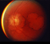
In adolescent- or young adult-onset disease (age <30 years), profound visual loss can be accompanied by minimal ophthalmoscopic findings and minimal ataxia [Thurtell et al 2009] (see Genotype-Phenotype Correlations). The onset of cone-rod dystrophy is often characterized by hemeralopia (inability to see clearly in bright light), photophobia (extreme sensitivity to light), decreased central visual acuity, and abnormalities in the tritan (blue-yellow) axis on detailed color vision testing [Miller et al 2009]. As cone function decreases over time, central visual acuity decreases to 20/200 (legally blind) and central scotomas develop; more prominent macular changes appear (see Figure 1), and all color discrimination is lost. Eventually all vision is lost.

Figure 1. . Funduscopic photo shows extreme macular degeneration of late-stage SCA7.

Figure 1.

Funduscopic photo shows extreme macular degeneration of late-stage SCA7.

Early signs of cone-rod dystrophy are subtle granular changes in the macula. Electroretinogram is consistently abnormal early in the disease course, showing a decrease in the photopic (cone) response initially, followed by a decrease in the scotopic (rod) response [Miller et al 2009].

In classic adult-onset disease (age >40 years), vision loss from retinal degeneration typically follows the onset of ataxia (sometimes many years to decades later) and gradually declines, seldom progressing to total blindness [Miller et al 2009].

Infantile- or Early Childhood-Onset SCA7

In infancy or early childhood disease, progression is always more rapid and aggressive than in adults. In infants, the clinical diagnosis may be elusive because ataxia and visual loss are not obvious; failure to thrive and loss of motor milestones may be the earliest findings. Other findings include progressive hypotonia, poor feeding, dysphagia, and congestive heart failure [Babovic-Vuksanovic et al 1998, Benton et al 1998]. Indeed, with rapid multisystem failure (including cerebellar and brain stem degeneration and other organ systems including lungs, heart, and kidneys), retinal degeneration and related vision loss may not be evident.

Affected infants usually die within months of initial presentation and never survive into early childhood [Ansorge et al 2004], a distinctly different clinical course from adult-onset SCA7, in which other organ system involvement does not occur (see Genotype-Phenotype Correlations).

Pathology. Neuronal loss, loss of myelinated fibers, and gliosis are observed in the cerebellum (especially Purkinje cells); the inferior olivary, dentate, and pontine nuclei; and to a lesser extent in the cerebral cortex, basal ganglia, thalamus, and midbrain [Rüb et al 2008, Seidel et al 2012].

Genotype-Phenotype Correlations

A correlation between CAG repeat sizes and disease severity exists: the longer the CAG repeat, the earlier the age of onset and the more severe and rapidly progressive the disease.

  • Infantile onset may be associated with CAG repeat sizes ranging from 200 to 400; however, technical limitations of genetic testing utilizing PCR amplification of the ATXN7CAG repeat region that often underestimate the repeat expansion size may report a CAG repeat size of fewer than 150.
  • Childhood onset is usually associated with CAG repeat sizes greater than 100.
  • Juvenile onset is often associated with CAG repeat sizes 60-100.

A correlation between CAG repeat size and initial clinical manifestation exists [Johansson et al 1998]:

  • CAG repeat sizes greater than 59 are typically associated with adolescent or young-adult onset (age <30 years) and visual impairment as the initial manifestation.
  • CAG repeat sizes smaller than 59 are often associated with adult onset (age >30 years) and cerebellar findings as the initial manifestation.

Despite observations correlating CAG repeat length with age of onset, disease severity, and course, CAG repeat size cannot provide sufficient predictive value for clinical prognosis within the classic adult-onset CAG repeat size range of 38 to 50 repeats [Andrew et al 1997].

Reports of pathogenic (age-related reduced-penetrance) repeats include the following:

Penetrance

See Genotype-Phenotype Correlations for CAG repeat sizes associated with age-related reduced penetrance.

Anticipation

In families with a pathogenic (full-penetrance) CAG repeat expansion, the repeat size tends to expand with transmission to successive generations, with more marked expansions seen in affected offspring of affected males [Gouw et al 1998]. This explains, at the genetic level, the marked anticipation seen in families with SCA7, now regarded as the most unstable of all CAG repeat disorders.

Anticipation in a family may be so dramatic that a child may be diagnosed with what is thought to be an unrelated neurodegenerative disease years before a parent or grandparent with pathogenic CAG repeat expansion becomes symptomatic [van de Warrenburg et al 2001, Ansorge et al 2004].

Repeat contraction has not been reported.

Nomenclature

Terms used in the past to designate SCA7 include olivopontocerebellar ataxia (OPCA) type III and ADCA type II.

Prevalence

The prevalence is fewer than 1:300,000. In several studies, SCA7 represented 2% of all SCAs [Filla et al 2000, Storey et al 2000].

SCA7 occurs predominantly in two racial population groups: northern Europeans and Africans. Indeed, SCA7 is the only repeat expansion disease, with the exception of Huntington disease-like 2 (HDL2), with a large number of affected individuals of African racial ancestry. For this reason, a substantial fraction of individuals with SCA7 in the United States are of African racial ancestry. Worldwide, SCA7 is seen in North America, Europe, Eurasia, Australia, South Africa, and South America.

As a result of a founder effect in Mexico dating back to the colonial era, a very large concentration of individuals with SCA7 have been ascertained in the state of Veracruz in Mexico, with well over 150 documented affected individuals.

Differential Diagnosis

While many of the neurologic and pathologic findings of the other spinocerebellar ataxias (SCAs) overlap with SCA7, retinal degeneration is the distinguishing feature of SCA7 (see Hereditary Ataxia Overview).

Table 3.

Disorders with Retinal Degeneration in the Differential Diagnosis of Spinocerebellar Ataxia Type 7

Gene(s)DisorderMOIEye FindingsNeurologic & Pathologic FindingsDistinguishing Features
CRX Cone-rod dystrophy 2 (OMIM 120970)ADImpaired color vision; central scotomaNo neurologic findingsNo neurologic findings
MT-ND1
MT-ND4
MT-ND6 1
Leber hereditary optic neuropathy MatImpaired color vision; central scotomaNo neurologic findingsUsually midlife presentation
OPA3 Costeff syndrome (3-methylglutaconic aciduria type 3)ARBilateral optic atrophyChorea, spastic paraparesis, mild ataxia
  • Optic atrophy in childhood (age <10 yrs)
  • Common in persons of Iraqi Jewish origin due to founder variant
WFS1
CISD2
Wolfram syndrome
(See WFS1 Wolfram Syndrome Spectrum Disorder.)
ARBilateral optic trophyAtaxia, diabetes mellitus/insipidus, hearing lossChildhood-onset diabetes mellitus & optic atrophy
1.

Three common mtDNA pathogenic variants in the listed genes account for 90%-95% of Leber hereditary optic neuropathy (LHON). Pathogenic variants in other mitochondrial genes (MT-ND2, MT-ND4L, and MT-ND5) are also known to be associated with LHON.

Management

Evaluations Following Initial Diagnosis

To establish the extent of disease and needs in an individual diagnosed with spinocerebellar ataxia type 7 (SCA7) of adolescent or adult onset, the evaluations summarized in Table 4 (if not performed as part of the evaluation that led to the diagnosis) are recommended.

Table 4.

Recommended Evaluations Following Initial Diagnosis of SCA7: Adolescent or Adult Onset

System/ConcernEvaluationComment
Neurologic Neurologist assessment for cerebellar motor dysfunction (gait & postural ataxia, dysmetria, dysdiadochokinesis, tremor, dysarthria, nystagmus, saccades & smooth pursuit)Use standardized scale to establish baseline for ataxia (SARA, ICARS, or BARS). 1
UMN &/or LMN dysfunction (weakness, spasticity, Babinski signs, hyperreflexia, amyotrophy, fasciculations)Since most exhibit some corticospinal tract involvement, comprehensive assessment of motor & sensory function recommended for all affected persons
Refer to neuromuscular clinic (OT/PT / rehab specialist).Assess gross motor & fine motor skills, gait, ambulation, need for adaptive devices, PT, &OT.
Ophthalmologic
involvement
Complete eye examIncl:
  • BCVA
  • Extraocular movement
  • Refractive error
  • Color vision testing
  • Full-field ERG
  • Spectral-domain OCT
Speech For those w/dysarthria: speech/language evalConsider involving certified practitioner of speech/language pathology.
Feeding For those w/frequent choking or severe dysphagia, assess:
  • Nutritional status;
  • Aspiration risk.
Consider involving gastroenterology/nutrition/feeding team.
Respiratory For those w/respiratory symptoms or muscular involvement: obtain pulmonary function tests.Consider involving pulmonary specialist / respiratory therapist.
Bladder
function
History of spastic bladder symptoms: urgency, frequency, difficulty voiding
  • Referral to urologist
  • Consider urodynamic eval.
Restless legs
syndrome
Obtain comprehensive history w/emphasis on triggering & relieving factors.Consider referral to specialist w/experience in caring for individuals w/SCA7.
Chronic pain A comprehensive history & physical & neurologic exam must be performed.Consider referral to specialist (e.g., pain clinic or pain service).
Cognitive/
Psychiatric
Assess for cognitive dysfunction assoc w/cerebellar cognitive & affective syndrome (executive function, language processing, visuospatial / visuoconstructional skills, emotion regulation)Consider use of:
  • CCAS scale 2 to evaluate cognitive & emotional involvement;
  • Psychiatrist, psychologist, neuropsychologist if needed.
Family support
& resources
Consider individual’s disease severity & ability to receive regular care & support from family.Assess need for:
  • Community or online resources;
  • Social work involvement for caregiver support;
  • Home nursing referral.
Genetic
counseling
By genetics professionals 3To inform affected persons & their families re nature, MOI, & implications of SCA7 to facilitate medical & personal decision making

BARS = Brief Ataxia Rating Scale; BCVA = best-corrected visual acuity; CCAS = cerebellar cognitive affective syndrome; ERG = electroretinogram; ICARS = International Co-operative Ataxia Rating Scale; LMN = lower motor neuron; MOI = mode of inheritance; OCT = optical coherence tomography; OT = occupational therapy; PT = physical therapy; SARA = Scale for the Assessment and Rating of Ataxia; UMN = upper motor neuron

1.
2.
3.

Medical geneticist, certified genetic counselor, or certified advanced genetic nurse

Treatment of Manifestations

Management of affected individuals remains supportive, as no known therapy to delay or halt the progression of the disease exists.

Table 5.

Treatment of Manifestations of SCA 7: Adolescent or Adult Onset

Manifestation/
Concern
TreatmentConsiderations/Other
Cerebellar ataxia PT/OT
  • PT (balance exercises, gait training, muscle strengthening) to maintain mobility & function 1
  • OT to optimize ADLs (incl use of adaptive devices, e.g., weighted eating utensils & dressing hooks)
  • Consider adaptive devices to maintain/improve independence in mobility (e.g., canes, walkers, motorized chairs).
  • Inpatient rehab w/OT/PT may improve ataxia & functional abilities. 2, 3
  • Weight control to avoid obesity
  • Home adaptations to prevent falls (e.g., grab bars, raised toilet seats) & improve mobility (e.g., ramps to accommodate motorized chairs)
Pharmacologic treatment
  • Therapies intended to ↓ symptoms work variably well in different individuals.
  • Most commonly used drugs: amantadine, buspirone, riluzole
TMSThis treatment modality is still being evaluated, w/most promising initial results obtained w/cerebellar repetitive TMS.
UMN involvement
(spasticity)
Pharmacologic treatmentConsider pharmacologic treatment of generalized spasticity w/oral medications (usually in this order due to the profile of side effects & better tolerance): baclofen, tizanidine, gabapentin, clonazepam, dantrolene sodium, diazepam
LMN involvement
(weakness)
Mainly supportiveBraces, orthotics, PT
Ophthalmologic
involvement
Use of low vision aidsConsultation w/agencies for visually impaired
Dysarthria Speech/language therapyConsider alternative communication methods as needed (e.g., writing pads & digital devices).
Dysphagia Feeding therapy programs to improve nutrition & dysphagia, & ↓ risk of aspirationVideo esophagram may help define best food consistency.
Weight Nutrition assessment
  • Consider nutritional & vitamin supplementation to meet dietary needs.
  • Avoid obesity (which can exacerbate difficulties with ambulation & mobility).
Bladder
dysfunction
Pharmacologic treatment
  • If physical rehab or biofeedback do not remedy problem, consider anticholinergic drugs for overactive bladder.
  • Anticholinergic agents are also indicated for neurogenic bladder.
  • Botulinum toxin injections should be reserved for severe or unresponsive bladder dysfunction.
Restless legs
syndrome
Pharmacologic treatmentLevodopa or dopamine agonist
Chronic pain Requires specialist eval & managementRefer to pain clinic or pain specialist.
Cognitive/
Psychiatric
Pharmacologic treatmentStandard treatment for psychiatric manifestations (e.g., depression, anxiety, & psychosis)
Psychotherapy / neuropsychological rehabConsider cognitive & behavioral therapy, incl Goal Management Training®4

ADLs = activities of daily living; LMN = lower motor neuron; OT = occupational therapy/therapist; PT = physical therapy/therapist; TMS = transcranial magnetic stimulation; UMN = upper motor neuron

1.
2.
3.
4.

Surveillance

Table 6.

Recommended Surveillance for Individuals with SCA7

System/ConcernEvaluationFrequency
Neurologic
  • Neurologic assessment for progression of ataxia; UMN or LMN signs; dystonia & parkinsonism; autonomic dysfunction
  • Monitor ataxia progression w/standardized scale (SARA, ICARS, or BARS). 1
Annually; more often for an acute exacerbation
Physiatry; OT/PT assessment of mobility; self-help skills as they relate to ataxia, spasticity, weakness
Dysarthria Assess need for alternative communication method or speech therapy.Per symptom progression
Dysphagia Assess aspiration risk & feeding methods.
Retinopathy Exam by ophthalmologist for evidence of cone-rod dystrophy: BCVA, color vision testing, visual field testing, ERGEvery 3-5 yrs or as needed based on concerns re visual acuity, visual field deficits, &/or color vision (as an indicator of cone function)
Cognitive/
Psychiatric
Evaluate mood, signs of psychosis, cognitive complaints to identify need for pharmacologic & psychotherapeutic interventions.Per symptom progression & development of psychiatric symptoms
Family support/
resources
Provide various options for affected persons & their families, ranging from joining patient support groups (e.g., National Ataxia Foundation, which has local chapters throughout the US) to social work consultation.Per symptom progression

BARS = Brief Ataxia Rating Scale; BCVA = best-corrected visual acuity; ERG = electroretinogram; ICARS = International Co-operative Ataxia Rating Scale; LMN = lower motor neuron; OT = occupational therapy; PT = physical therapy; SARA = Scale for the Assessment and Rating of Ataxia; UMN = upper motor neuron

1.

Agents/Circumstances to Avoid

Avoid drinking alcoholic beverages, as alcohol intake can further impair cerebellar function, especially if excessive.

Avoid foods identified by a registered dietician as potentially causing dizziness or disorientation.

Evaluation of Relatives at Risk

See Genetic Counseling for issues related to testing of at-risk relatives for genetic counseling purposes.

Therapies Under Investigation

Ongoing clinical trials for SCA7 include a study of:

  • Troriluzole in adults as a treatment for ataxia in the United States (ClinicalTrials.gov: NCT03701399);
  • Riluzole in adults as a treatment for ataxia in Italy (ClinicalTrials.gov: NCT03660917).

Ionis Pharmaceuticals™ is developing an antisense oligonucleotide for dosage reduction of ataxin-7 in the retina and brain, as a preclinical trial of this strategy was found to be an effective treatment for retinal degeneration in an SCA7 mouse model [Niu et al 2018].

Search ClinicalTrials.gov in the US and EU Clinical Trials Register in Europe for access to information on clinical studies for a wide range of diseases and conditions.

Genetic Counseling

Genetic counseling is the process of providing individuals and families with information on the nature, mode(s) of inheritance, and implications of genetic disorders to help them make informed medical and personal decisions. The following section deals with genetic risk assessment and the use of family history and genetic testing to clarify genetic status for family members; it is not meant to address all personal, cultural, or ethical issues that may arise or to substitute for consultation with a genetics professional. —ED.

Mode of Inheritance

Spinocerebellar ataxia type 7 (SCA7) is inherited in an autosomal dominant manner.

Risk to Family Members

Parents of a proband

  • Most individuals diagnosed with SCA7 have an affected parent. Note: Anticipation in a family may be so dramatic that a child may be diagnosed with SCA7 years before a parent or grandparent with pathogenic CAG repeat expansion becomes symptomatic [van de Warrenburg et al 2001, Ansorge et al 2004].
  • A proband with SCA7 may have the disorder as the result of expansion of a pathogenic reduced-penetrance CAG repeat (34-36 CAG repeats) or a mutable normal CAG repeat (28-33 CAG repeats) inherited from an unaffected parent.
  • If neither of the parents of the proband is known to have SCA7, recommendations for the evaluation of parents include physical examination and consideration of ATXN7 molecular genetic testing.
  • The family history of some individuals diagnosed with SCA7 may appear to be negative because of failure to recognize the disorder in family members, early death of the parent before the onset of manifestations, or late onset of the disease in the affected parent. Therefore, an apparently negative family history cannot be confirmed unless appropriate molecular genetic testing has been performed on the parents of the proband.

Sibs of a proband. The risk to the sibs of a proband depends on the clinical/genetic status of the parents:

  • If a parent of the proband is affected and/or is known to have an abnormal CAG repeat, the risk to each sib of inheriting the CAG repeat expansion is 50%. Pathogenic full-penetrance CAG repeat expansions tend to expand on transmission from parent to offspring (more marked expansions are seen when the transmitting parent is male) and often result in an earlier age of onset and more severe disease manifestations in offspring (see Anticipation).
  • Clinical presentation in sibs who inherit an abnormal CAG repeat usually correlates with their CAG repeat size: the longer the CAG repeat, the earlier the age of onset and the more severe and rapidly progressive the disease (see Genotype-Phenotype Correlations).
  • If the parents of a proband are clinically unaffected but their genetic status is unknown, sibs are still presumed to be at increased risk for SCA7 because of the possibility of late onset of SCA7 in a heterozygous parent.

Offspring of a proband

  • Each child of an affected individual has a 50% chance of inheriting the CAG repeat expansion.
  • Pathogenic full-penetrance CAG repeat expansions tend to expand on transmission from parent to offspring (more marked expansions are seen when the transmitting parent is male) and often result in an earlier age of onset and more severe disease manifestations in offspring (see Anticipation).

Other family members. The risk to other family members depends on the genetic status of the proband's parents: if a parent has the CAG repeat expansion, the parent's family members are at risk.

Related Genetic Counseling Issues

Note: If neither parent of a proband with SCA7 has a CAG repeat expansion, nonmedical explanations including alternate paternity or maternity (e.g., with assisted reproduction) and undisclosed adoption could also be explored.

Family planning

  • The optimal time for determination of genetic risk and discussion of the availability of prenatal/preimplantation genetic testing is before pregnancy.
  • It is appropriate to offer genetic counseling (including discussion of potential risks to offspring and reproductive options) to young adults who are affected or at risk.

At-risk individuals. The age of onset, severity, specific manifestations, and progression of the disease are variable and cannot be reliably predicted by the family history or molecular genetic testing.

Predictive testing (i.e., testing of asymptomatic at-risk individuals)

  • Predictive testing for at-risk relatives is possible once molecular genetic testing has identified a CAG repeat expansion in an affected family member.
  • Potential consequences of such testing (including but not limited to socioeconomic changes and the need for long-term follow up and evaluation arrangements for individuals with a positive test result) as well as the capabilities and limitations of predictive testing should be discussed in the context of formal genetic counseling prior to testing.

Predictive testing in minors (i.e., testing of asymptomatic at-risk individuals age <18 years)

  • For asymptomatic minors at risk for adult-onset conditions for which early treatment would have no beneficial effect on disease morbidity and mortality, predictive genetic testing is considered inappropriate, primarily because it negates the autonomy of the child with no compelling benefit. Further, concern exists regarding the potential unhealthy adverse effects that such information may have on family dynamics, the risk of discrimination and stigmatization in the future, and the anxiety that such information may cause.
  • For more information, see the National Society of Genetic Counselors position statement on genetic testing of minors for adult-onset conditions and the American Academy of Pediatrics and American College of Medical Genetics and Genomics policy statement: ethical and policy issues in genetic testing and screening of children.

In a family with an established diagnosis of SCA7, it is appropriate to consider testing of symptomatic individuals regardless of age.

Prenatal Testing and Preimplantation Genetic Testing

Once the ATXN7 CAG repeat expansion has been identified in an affected family member, prenatal testing for a pregnancy at increased risk and preimplantation genetic testing are possible. (Note: While the prenatal finding of a CAG repeat expansion cannot be used to accurately predict onset, severity, specific manifestation, or rate of progression of SCA7, disease-causing alleles longer than 59 CAG repeats usually result in onset before age 30 years.)

Differences in perspective may exist among medical professionals and within families regarding the use of prenatal testing. While most centers would consider use of prenatal testing to be a personal decision, discussion of these issues may be helpful.

Resources

GeneReviews staff has selected the following disease-specific and/or umbrella support organizations and/or registries for the benefit of individuals with this disorder and their families. GeneReviews is not responsible for the information provided by other organizations. For information on selection criteria, click here.

  • NCBI Genes and Disease
  • Ataxia UK
    United Kingdom
    Phone: 0800 995 6037; +44 (0) 20 7582 1444 (from abroad)
    Email: help@ataxia.org.uk
  • euro-ATAXIA (European Federation of Hereditary Ataxias)
    United Kingdom
    Email: lporter@ataxia.org.uk
  • National Ataxia Foundation
    Phone: 763-553-0020
    Fax: 763-553-0167
    Email: naf@ataxia.org
  • Spanish Ataxia Federation (FEDAES)
    Spain
    Phone: 601 037 982
    Email: info@fedaes.org
  • CoRDS Registry
    Sanford Research
    Phone: 605-312-6300

Molecular Genetics

Information in the Molecular Genetics and OMIM tables may differ from that elsewhere in the GeneReview: tables may contain more recent information. —ED.

Table A.

Spinocerebellar Ataxia Type 7: Genes and Databases

GeneChromosome LocusProteinLocus-Specific DatabasesHGMDClinVar
ATXN7 3p14​.1 Ataxin-7 ATXN7 database ATXN7 ATXN7

Data are compiled from the following standard references: gene from HGNC; chromosome locus from OMIM; protein from UniProt. For a description of databases (Locus Specific, HGMD, ClinVar) to which links are provided, click here.

Table B.

OMIM Entries for Spinocerebellar Ataxia Type 7 (View All in OMIM)

164500SPINOCEREBELLAR ATAXIA 7; SCA7
607640ATAXIN 7; ATXN7

Molecular Pathogenesis

ATXN7 encodes ataxin-7, a predominantly nuclear protein that shuttles between the nucleus and cytoplasm. Ataxin-7 is a core component of a transcription co-activator complex called STAGA [Garden & LaSpada 2008]. Ataxin-7 also has a cytoplasmic role in stabilizing microtubules [Nakamura et al 2012]. The normal distribution of ataxin-7 in human brain and retina has been described [Cancel et al 2000].

Mechanism of disease causation. Gain of function as a result of a CAG expansion in ATXN7

Table 7.

ATXN7 Technical Considerations

Technical IssueComment [Reference]
Sequence of repeatCAG
Methods to detect expanded allele (See Table 8.)PCR w/fragment analysis, triplet-primed PCR, & Southern blotting
Somatic instabilityAlleles w/abnormal number of CAG repeats may display somatic instability of the repeat, appearing as "smeared" expanded alleles w/multiple distinct expansion alleles on PCR or Southern blot analysis [Gu et al 2000, Katagiri et al 2015].
Germline instabilityThe CAG repeat size tends to expand w/transmission to successive generations, w/more marked expansions seen in affected offspring of affected males [Gouw et al 1998].

Methods to characterize ATXN7 CAG repeats. Because of the technical challenges of detecting and sizing ATXN7 CAG repeat expansions (see Table 7), multiple methods may be needed to rule out or detect an expanded allele (see Table 8). Repeats up to about 100 CAG may be detected by traditional PCR. However, detection of apparent homozygosity for a normal CAG repeat does not rule out the presence of an expanded CAG repeat; thus, testing by triplet-primed PCR (TP-PCR) or Southern blotting is required.

In addition, somatic and germline instability of expanded repeats must be considered.

Table 8.

Methods to Characterize ATXN7 CAG Repeats

Interpretation of CAG Repeat NumberExpected Results by Method
Conventional PCRTriplet-Primed PCR 1Expanded Repeat Analysis 2
Normal, uncertain, mutable normal, reduced penetrance: <19-36 rptsDetected 3See footnote 1.Expansions detected, & repeat size can be approximated. 7, 8
Pathogenic (full penetrance): 37-~460 rptsDetected up to ~100 CAG 4Expansions detected, but repeat size cannot be determined. 5, 6
1.

The design of a triplet-primed PCR (TP-PCR) assay may include conventional PCR primers to size normal repeats and detect expanded repeats in a single assay. The TP-PCR assay itself does not determine repeat size, even alleles in the normal range.

2.

Methods to detect and approximate the size of expanded repeats include long-range PCR sized by gel electrophoresis and Southern blotting. The upper limit of repeat size detected will vary by assay design, laboratory, sample, and/or patient as a result of competition by the normal allele during amplification.

3.

Detection of an apparently homozygous repeat does not rule out the presence of an expanded CAG repeat; thus, testing by TP-PCR or expanded repeat analysis is required to detect a repeat expansion.

4.

The upper limit of repeat size detected will vary by assay design, laboratory, sample, and/or patient as a result of competition from the normal allele during amplification.

5.

TP-PCR for the CAG repeat expansion has been described [Cagnoli et al 2006].

6.

Repeats at the lower end of this range may not show the characteristic stutter pattern that indicates an expanded repeat [Cagnoli et al 2006].

7.

Southern blotting for the CAG repeat expansion has been described [Benton et al 1998].

8.

Precise sizing of repeats is not necessary, as clinical utility for determining the exact repeat number has not been demonstrated.

Table 9.

Notable ATXN7 Pathogenic Variants

Reference SequencesDNA Nucleotide
Change
Predicted
Protein Change
Repeat Range [Reference]
NM_000333​.3
NP_000324​.1
c.89_91AGC[7_27]p.Gln38[7_27]Normal [David et al 1997, Benton et al 1998]
c.89_91AGC[28_33]p.Gln38[28_33]Mutable normal [Lebre et al 2003, Mittal et al 2005]
c.89_91AGC[34_36]p.Gln38[34_36]Pathogenic reduced penetrance [David et al 1998, Koob et al 1998, Stevanin et al 1998, Nardacchione et al 1999]
c.89_91AGC[37_~460]p.Gln38[37_~460]Pathogenic full penetrance [David et al 1997, Benton et al 1998, Michalik et al 2004]

Variants listed in the table have been provided by the author. GeneReviews staff have not independently verified the classification of variants.

GeneReviews follows the standard naming conventions of the Human Genome Variation Society (varnomen​.hgvs.org). See Quick Reference for an explanation of nomenclature.

References

Published Guidelines / Consensus Statements

  • Committee on Bioethics, Committee on Genetics, and American College of Medical Genetics and Genomics Social, Ethical, Legal Issues Committee. Ethical and policy issues in genetic testing and screening of children. Available online. 2013. Accessed 6-10-22.
  • National Society of Genetic Counselors. Position statement on genetic testing of minors for adult-onset conditions. Available online. 2018. Accessed 6-10-22.

Literature Cited

  • Ahn JK, Seo JM, Chung H, Yu HG. Anatomical and functional characteristics in atrophic maculopathy associated with spinocerebellar ataxia type 7. Am J Ophthalmol. 2005;139:923–5. [PubMed: 15860307]
  • Aleman TS, Cideciyan AV, Volpe NJ, Stevanin G, Brice A, Jacobson SG. Spinocerebellar ataxia type 7 (SCA7) shows a cone-rod dystrophy phenotype. Exp Eye Res. 2002;74:737–45. [PubMed: 12126946]
  • Andrew SE, Goldberg YP, Hayden MR. Rethinking genotype and phenotype correlations in polyglutamine expansion disorders. Hum Mol Genet. 1997;6:2005–10. [PubMed: 9328463]
  • Ansorge O, Giunti P, Michalik A, Van Broeckhoven C, Harding B, Wood N, Scaravilli F. Ataxin-7 aggregation and ubiquitination in infantile SCA7 with 180 CAG repeats. Ann Neurol. 2004;56:448–52. [PubMed: 15349877]
  • Babovic-Vuksanovic D, Snow K, Patterson MC, Michels VV. Spinocerebellar ataxia type 2 (SCA 2) in an infant with extreme CAG repeat expansion. Am J Med Genet. 1998;79:383–7. [PubMed: 9779806]
  • Benton CS, de Silva R, Rutledge SL, Bohlega S, Ashizawa T, Zoghbi HY. Molecular and clinical studies in SCA-7 define a broad clinical spectrum and the infantile phenotype. Neurology. 1998;51:1081–6. [PubMed: 9781533]
  • Bürk K, Sival DA. Scales for the clinical evaluation of cerebellar disorders. Handb Clin Neurol. 2018;154:329–39. [PubMed: 29903450]
  • Cagnoli C, Stevanin G, Michielotto C, Gerbino Promis G, Brussino A, Pappi P, Durr A, Dragone E, Viemont M, Gellera C, Brice A, Migone N, Brusco A. Large pathogenic expansions in the SCA2 and SCA7 genes can be detected by fluorescent repeat-primed polymerase chain reaction assay. J Mol Diagn. 2006;8:128–32. [PMC free article: PMC1867568] [PubMed: 16436644]
  • Cancel G, Duyckaerts C, Holmberg M, Zander C, Yvert G, Lebre AS, Ruberg M, Faucheux B, Agid Y, Hirsch E, Brice A. Distribution of ataxin-7 in normal human brain and retina. Brain. 2000;123:2519–30. [PubMed: 11099453]
  • David G, Abbas N, Stevanin G, Dürr A, Yvert G, Cancel G, Weber C, Imbert G, Saudou F, Antoniou E, Drabkin H, Gemmill R, Giunti P, Benomar A, Wood N, Ruberg M, Agid Y, Mandel JL, Brice A. Cloning of the SCA7 gene reveals a highly unstable CAG repeat expansion. Nat Genet. 1997;17:65–70. [PubMed: 9288099]
  • David G, Dürr A, Stevanin G, Cancel G, Abbas N, Benomar A, Belal S, Lebre AS, Abada-Bendib M, Grid D, Holmberg M, Yahyaoui M, Hentati F, Chkili T, Agid Y, Brice A. Molecular and clinical correlations in autosomal dominant cerebellar ataxia with progressive macular dystrophy (SCA7). Hum Mol Genet. 1998;7:165–70. [PubMed: 9425222]
  • Filla A, Mariotti C, Caruso G, Coppola G, Cocozza S, Castaldo I, Calabrese O, Salvatore E, De Michele G, Riggio MC, Pareyson D, Gellera C, Di Donato S. Relative frequencies of CAG expansions in spinocerebellar ataxia and dentatorubropallidoluysian atrophy in 116 Italian families. Eur Neurol. 2000;44:31–6. [PubMed: 10894992]
  • Garden GA, La Spada AR. Molecular pathogenesis and cellular pathology of spinocerebellar ataxia type 7 neurodegeneration. Cerebellum. 2008;7:138–49. [PMC free article: PMC4195584] [PubMed: 18418675]
  • Giunti P, Stevanin G, Worth PF, David G, Brice A, Wood NW. Molecular and clinical study of 18 families with ADCA type II: evidence for genetic heterogeneity and de novo mutation. Am J Hum Genet. 1999;64:1594–603. [PMC free article: PMC1377902] [PubMed: 10330346]
  • Gouw LG, Castañeda MA, McKenna CK, Digre KB, Pulst SM, Perlman S, Lee MS, Gomez C, Fischbeck K, Gagnon D, Storey E, Bird T, Jeri FR, Ptácek LJ. Analysis of the dynamic mutation in the SCA7 gene shows marked parental effects on CAG repeat transmission. Hum Mol Genet. 1998;7:525–32. [PubMed: 9467013]
  • Gu W, Wang Y, Liu X, Zhou B, Zhou Y, Wang G. Molecular and clinical study of spinocerebellar ataxia type 7 in Chinese kindreds. Arch Neurol. 2000;57:1513–8. [PubMed: 11030806]
  • Hoche F, Guell X, Vangel MG, Sherman JC, Schmahmann JD. The cerebellar cognitive affective/Schmahmann syndrome scale. Brain. 2018;141:248–70. [PMC free article: PMC5837248] [PubMed: 29206893]
  • Hugosson T, Gränse L, Ponjavic V, Andréasson S. Macular dysfunction and morphology in spinocerebellar ataxia type 7 (SCA 7). Ophthalmic Genet. 2009;30:1–6. [PubMed: 19172503]
  • Johansson J, Forsgren L, Sandgren O, Brice A, Holmgren G, Holmberg M. Expanded CAG repeats in Swedish spinocerebellar ataxia type 7 (SCA7) patients: effect of CAG repeat length on the clinical manifestation. Hum Mol Genet. 1998;7:171–6. [PubMed: 9425223]
  • Katagiri S, Hayashi T, Takeuchi T, Yamada H, Gekka T, Kawabe K, Kurita A, Tsuneoka H. Somatic instability of expanded CAG repeats of ATXN7 in Japanese patients with spinocerebellar ataxia type 7. Doc Ophthalmol. 2015;130:189–95. [PubMed: 25643591]
  • Koob MD, Benzow KA, Bird TD, Day JW, Moseley ML, Ranum LP. Rapid cloning of expanded trinucleotide repeat sequences from genomic DNA. Nat Genet. 1998;18:72–5. [PubMed: 9425905]
  • Lebre AS, Stevanin G, Brice A. Spinocerebellar ataxia 7 (SCA7). In: Pulst SM, ed. Genetics of Movement Disorders. San Diego, CA: Academic Press; 2003:85-94.
  • Martineau L, Noreau A, Dupré N. Therapies for ataxias. Curr Treat Options Neurol. 2014;16:300. [PubMed: 24832479]
  • Michalik A, Martin JJ, Van Broeckhoven C. Spinocerebellar ataxia type 7 associated with pigmentary retinal dystrophy. Eur J Hum Genet. 2004;12:2–15. [PubMed: 14571264]
  • Miller RC, Tewari A, Miller JA, Garbern J, Van Stavern GP. Neuro-ophthalmologic features of spinocerebellar ataxia type 7. J Neuroophthalmol. 2009;29:180–6. [PubMed: 19726938]
  • Mittal U, Roy S, Jain S, Srivastava AK, Mukerji M. Post-zygotic de novo trinucleotide repeat expansion at spinocerebellar ataxia type 7 locus: evidence from an Indian family. J Hum Genet. 2005;50:155–7. [PubMed: 15750685]
  • Nakamura Y, Tagawa K, Oka T, Sasabe T, Ito H, Shiwaku H, La Spada AR, Okazawa H. Ataxin-7 associates with microtubules and stabilizes the cytoskeletal network. Hum Mol Genet. 2012;21:1099–110. [PMC free article: PMC3277310] [PubMed: 22100762]
  • Nardacchione A, Orsi L, Brusco A, Franco A, Grosso E, Dragone E, Mortara P, Schiffer D, De Marchi M. Definition of the smallest pathological CAG expansion in SCA7. Clin Genet. 1999;56:232–4. [PubMed: 10563484]
  • Niu C, Prakash TP, Kim A, Quach JL, Huryn LA, Yang Y, Lopez E, Jazayeri A, Hung G, Sopher BL, Brooks BP, Swayze EE, Bennett CF, La Spada AR. Delivery of antisense oligonucleotides into the vitreous humor yields effective suppression of misfolded protein toxicity in SCA7 retinal degeneration. Sci Transl Med. 2018;10:eaap8677. [PMC free article: PMC6411060] [PubMed: 30381411]
  • Rüb U, Brunt ER, Seidel K, Gierga K, Mooy CM, Kettner M, Van Broeckhoven C, Bechmann I, La Spada AR, Schöls L, den Dunnen W, de Vos RA, Deller T. Spinocerebellar ataxia type 7 (SCA7): widespread brain damage in an adult-onset patient with progressive visual impairments in comparison with an adult-onset patient without visual impairments. Neuropathol Appl Neurobiol. 2008;34:155–68. [PubMed: 17971076]
  • Ruffieux N, Colombo F, Gentaz E, Annoni JM, Chouiter L, Roulin Hefti S, Ruffieux A, Bihl T. Successful neuropsychological rehabilitation in a patient with cerebellar cognitive affective syndrome. Appl Neuropsychol Child. 2017;6:180–8. [PubMed: 27049666]
  • Seidel K, Siswanto S, Brunt ER, den Dunnen W, Korf HW, Rüb U. Brain pathology of spinocerebellar ataxias. Acta Neuropathol. 2012;124:1–21. [PubMed: 22684686]
  • Sokolovsky N, Cook A, Hunt H, Giunti P, Cipolotti L. A preliminary characterisation of cognition and social cognition in spinocerebellar ataxia types 2, 1, and 7. Behav Neurol. 2010;23:17–29. [PMC free article: PMC5434399] [PubMed: 20714058]
  • Stevanin G, Giunti P, Belal GD, Dürr A, Ruberg M, Wood N, Brice A. De novo expansion of intermediate alleles in spinocerebellar ataxia 7. Hum Mol Genet. 1998;7:1809–13. [PubMed: 9736784]
  • Storey E, du Sart D, Shaw JH, Lorentzos P, Kelly L, McKinley Gardner RJ, Forrest SM, Biros I, Nicholson GA. Frequency of spinocerebellar ataxia types 1, 2, 3, 6, and 7 in Australian patients with spinocerebellar ataxia. Am J Med Genet. 2000;95:351–7. [PubMed: 11186889]
  • Thurtell MJ, Fraser JA, Bala E, Tomsak RL, Biousse V, Leigh RJ, Newman NJ. Two patients with spinocerebellar ataxia type 7 presenting with profound binocular visual loss yet minimal ophthalmoscopic findings. J Neuroophthalmol. 2009;29:187–91. [PMC free article: PMC2987707] [PubMed: 19726939]
  • To KW, Adamian M, Jakobiec FA, Berson EL. Olivopontocerebellar atrophy with retinal degeneration. An electroretinographic and histopathologic investigation. Ophthalmology. 1993;100:15–23. [PubMed: 8433819]
  • van de Warrenburg BP, Frenken CW, Ausems MG, Kleefstra T, Sinke RJ, Knoers NV, Kremer HP. Striking anticipation in spinocerebellar ataxia type 7: the infantile phenotype. J Neurol. 2001;248:911–4. [PubMed: 11697534]
  • van de Warrenburg BP, van Gaalen J, Boesch S, Burgunder JM, Durr A, Giunti P, Klockgether T, Mariotti C, Pandolfo M, Riess O. EFNS/ENS consensus on the diagnosis and management of chronic ataxias in adulthood. Eur J Neurol. 2014;21:552–62. [PubMed: 24418350]
  • Zesiewicz TA, Wilmot G, Kuo SH, Perlman S, Greenstein PE, Ying SH, Ashizawa T, Subramony SH, Schmahmann JD, Figueroa KP, Mizusawa H, Schöls L, Shaw JD, Dubinsky RM, Armstrong MJ, Gronseth GS, Sullivan KL. Comprehensive systematic review summary: treatment of cerebellar motor dysfunction and ataxia: Report of the Guideline Development, Dissemination, and Implementation Subcommittee of the American Academy of Neurology. Neurology. 2018;90:464–71. [PMC free article: PMC5863491] [PubMed: 29440566]

Chapter Notes

Author History

Thomas D Bird, MD; University of Washington (2007-2012)
Gwenn Garden, MD, PhD; University of Washington (2012-2020)
Launce G-C Gouw, MD, PhD; University of Utah School of Medicine (1998-2007)
Albert R La Spada, MD, PhD (2007-2012; 2020-present)
Roberta A Pagon, MD; University of Washington (2007-2012)
Louis J Ptacek, MD; University of California, San Francisco (1998-2007)

Revision History

  • 23 July 2020 (bp) Comprehensive update posted live
  • 20 December 2012 (me) Comprehensive update posted live
  • 6 September 2007 (tb) Revision: Natural History (Clinical Description)
  • 9 February 2007 (me) Comprehensive update posted live
  • 11 December 2003 (me) Comprehensive update posted live
  • 20 June 2001 (me) Comprehensive update posted live
  • 27 August 1998 (pb) Review posted live
  • 1 June 1998 (lg) Original submission
Copyright © 1993-2024, University of Washington, Seattle. GeneReviews is a registered trademark of the University of Washington, Seattle. All rights reserved.

GeneReviews® chapters are owned by the University of Washington. Permission is hereby granted to reproduce, distribute, and translate copies of content materials for noncommercial research purposes only, provided that (i) credit for source (http://www.genereviews.org/) and copyright (© 1993-2024 University of Washington) are included with each copy; (ii) a link to the original material is provided whenever the material is published elsewhere on the Web; and (iii) reproducers, distributors, and/or translators comply with the GeneReviews® Copyright Notice and Usage Disclaimer. No further modifications are allowed. For clarity, excerpts of GeneReviews chapters for use in lab reports and clinic notes are a permitted use.

For more information, see the GeneReviews® Copyright Notice and Usage Disclaimer.

For questions regarding permissions or whether a specified use is allowed, contact: ude.wu@tssamda.

Bookshelf ID: NBK1256PMID: 20301433

Views

Tests in GTR by Gene

Related information

  • MedGen
    Related information in MedGen
  • OMIM
    Related OMIM records
  • PMC
    PubMed Central citations
  • PubMed
    Links to PubMed
  • Gene
    Locus Links

Similar articles in PubMed

See reviews...See all...

Recent Activity

Your browsing activity is empty.

Activity recording is turned off.

Turn recording back on

See more...