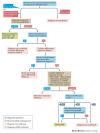
U.S. flag

An official website of the United States government

NCBI Bookshelf. A service of the National Library of Medicine, National Institutes of Health.

Adam MP, Feldman J, Mirzaa GM, et al., editors. GeneReviews® [Internet]. Seattle (WA): University of Washington, Seattle; 1993-2024.

Cover of GeneReviews®

GeneReviews® [Internet].

Show details

Silver-Russell Syndrome

Synonym: Russell-Silver Syndrome

, MD, , MD, and , MD, PhD.

Author Information and Affiliations

Initial Posting: ; Last Update: May 9, 2024.

Estimated reading time: 58 minutes

Summary

Clinical characteristics.

Silver-Russell Syndrome (SRS) is typically characterized by gestational growth restriction resulting in affected individuals being born small for gestational age, with relative macrocephaly at birth (head circumference ≥1.5 standard deviations [SD] above birth weight and/or length), prominent forehead with frontal bossing, and frequently body asymmetry. This is typically followed by postnatal growth failure, and in some cases progressive limb length discrepancy and feeding difficulties. Additional clinical features include triangular facies, fifth finger clinodactyly, and micrognathia with narrow chin. Except for the limb length asymmetry, growth failure is proportionate and head growth typically normal. The average adult height in untreated individuals is ~3.1±1.4 SD below the mean. The Netchine-Harbison Clinical Scoring System (NH-CSS) is a sensitive diagnostic scoring system. Clinical diagnosis can be established in an individual who meets at least four of the NH-CSS clinical criteria – prominent forehead/frontal bossing and relative macrocephaly at birth plus two additional findings – and in whom other disorders have been ruled out.

Diagnosis/testing.

SRS is a genetically heterogeneous condition. Genetic testing confirms clinical diagnosis in approximately 60% of affected individuals. Hypomethylation of the imprinting control region 1 (ICR1) at 11p15.5 causes SRS in 35%-67% of individuals, and maternal uniparental disomy of chromosome 7 (upd(7)mat) causes SRS in 7%-10% of individuals. There are a small number of individuals with SRS who have duplications, deletions, or translocations involving the imprinting centers at 11p15.5 or duplications, deletions, or translocations involving chromosome 7. Rarely, affected individuals with pathogenic variants in CDKN1C, IGF2, PLAG1, and HMGA2 have been described. However, approximately 30%-40% of individuals who meet NH-CSS clinical criteria for SRS have negative molecular and/or cytogenetic testing.

Management.

Treatment of manifestations: Multidisciplinary follow up and early specific intervention are necessary for optimal management of affected individuals, including early referral to an endocrinologist. Treatment may include growth hormone therapy. Hypoglycemia should be prevented or aggressively managed. Strategies for feeding issues include nutritional and caloric supplementation, medication for gastroesophageal reflux, therapy for oral motor problems and oral aversion, cyproheptadine for appetite stimulation, and enteral tube feeding as needed. Lower limb length discrepancy exceeding 2 cm requires intervention. In the majority of affected older children, distraction osteogenesis is recommended. Severe micrognathia or cleft palate should be managed by a multidisciplinary craniofacial team. Males with cryptorchidism or hypospadias should be referred to a urologist. Males with micropenis and females with internal genitourinary anomalies benefit from a referral to a multidisciplinary disorders of sex development (DSD) center. Physical, occupational, speech, and language therapy with an individualized education plan are used to treat developmental delays. Psychological counseling can be used as needed to address psychosocial and body image issues.

Surveillance: Monitoring of growth velocity, blood glucose concentration, and urine ketones for hypoglycemia in infants and as needed in older children; evaluation of nutritional status and oral intake at each visit as well as managing tube feedings if needed; limb length assessment at each well child visit in early childhood for evidence of asymmetric growth; evaluation for scoliosis, signs of precocious puberty, genitourinary issues, dental crowding and malocclusion, and speech-language development at each visit.

Agents/circumstances to avoid: Prolonged fasting in infants and young children because of the risk for hypoglycemia; elective surgery whenever possible due to risk of hypoglycemia, hypothermia, difficult healing, and difficult intubation.

Genetic counseling.

Risk to family members: In most families, a proband with SRS represents a simplex case (a single affected family member) and has SRS as a result of an apparent de novo epigenetic or genetic alteration. While the majority of families are presumed to have a very low recurrence risk, SRS can occur as the result of a genetic alteration associated with up to a 50% recurrence risk depending on the nature of the genetic alteration and the sex of the transmitting parent. Rare familial cases of SRS have been reported with several underlying mechanisms, including maternally inherited 11p15 duplications, maternally inherited CDKN1C gain-of-function pathogenic variants, paternally inherited IGF2 loss-of-function pathogenic variants, paternally inherited small deletions close to the boundaries of the ICR1, and paternally or maternally inherited deletions and intragenic pathogenic variants involving PLAG1 or HMGA2. Reliable SRS recurrence risk assessment therefore requires identification of the causative genetic mechanism in the proband.

Prenatal testing: Reliable prenatal testing for loss of paternal methylation at the 11p1.5 ICR1 H19/IGF2 region is not possible. Prenatal testing for the following SRS-related genetic mechanisms is possible provided the genetic mechanism has been demonstrated to be causative in an affected family member: upd(7)mat, SRS-related chromosomal abnormalities, intragenic CDKN1C, IGF2, HMGA2, or PLAG1 pathogenic variants, and deletions involving HMGA2 or PLAG1. The prenatal finding of a genetic alteration consistent with SRS cannot be used to reliably predict clinical outcome because children with SRS demonstrate varying responses to growth hormone, variable late catch-up growth, and variable developmental outcomes.

Diagnosis

Consensus clinical diagnostic criteria for Silver-Russell syndrome (SRS) have been published [Azzi et al 2015].

Suggestive Findings

SRS should be suspected in a proband with at least four of the following NH-CSS clinical criteria [Azzi et al 2015]:

  • Small for gestational age (birth weight and/or length two or more standard deviations [SD] below the mean for gestational age)
  • Postnatal growth failure (length/height two or more SD below the mean for age and sex at age 24 months)
  • Relative macrocephaly at birth (head circumference more than 1.5 SD above birth weight and/or length)
  • Frontal bossing or prominent forehead (forehead projecting beyond the facial plane on a side view at age one to three years)
  • Body asymmetry (limb length discrepancy ≥0.5 cm, or <0.5 cm with ≥2 other asymmetric body parts)
  • Feeding difficulties, or body mass index two or more SD below the mean at age two years, or current use of a feeding tube or cyproheptadine for appetite stimulation

Rarely, individuals meeting three of the six criteria have had positive molecular confirmation of SRS.

Establishing the Diagnosis

The clinical diagnosis of SRS can be established in a proband based on clinical diagnostic criteria [Azzi et al 2015], or the molecular diagnosis can be established in a proband with suggestive findings and abnormal molecular genetic testing consistent with one of several molecular mechanisms (see Table 1 and Molecular Pathogenesis).

Note: Molecular genetic testing is recommended in all individuals with suspected SRS to provide accurate recurrence risk information.

Clinical Diagnosis

SRS is an etiologically heterogeneous condition. In the international consensus statement for the diagnosis and management of SRS [Wakeling et al 2017], the NH-CSS was selected as the most sensitive of several compared clinical diagnostic scoring systems [Netchine et al 2007, Azzi et al 2015].

The clinical diagnosis of SRS can be established in a proband with at least four of the six NH-CSS clinical diagnostic criteria (see Suggestive Findings).

  • Two of the criteria must be relative macrocephaly at birth and frontal bossing or prominent forehead.
  • Other disorders with growth restriction must be ruled out either based on clinical features or molecular testing [Wakeling et al 2017] (see Differential Diagnosis).

Note: In older children and adults, frontal bossing or a prominent forehead may disappear. Therefore, a clinical diagnosis of SRS in older children and adults may require assessment of profile photographs from age two years or younger.

Molecular Diagnosis

The molecular diagnosis of SRS can be established in a proband with one of the following identified on molecular genetic testing (see Table 1 and Molecular Pathogenesis):

An international expert consensus algorithm for the molecular investigation of SRS has been published [Wakeling et al 2017], including first-tier, second-tier, and other molecular diagnostic testing approaches (see Figure 1). Third-tier testing using comprehensive genomic approaches has been proposed by the authors of this GeneReview and adapted from Wakeling et al [2017].

Figure 1.

Figure 1.

Flowchart for investigation and diagnosis of Silver-Russell syndrome Adapted from Wakeling et al [2017]

  • First-tier testing. Methylation analysis of 11p15.5 imprinting control regions (ICR1/ICR2) and upd(7)mat studies performed simultaneously.
    Note: (1) Detection of an abnormality is dependent on the mechanism of disease and testing methodology used (see Table 1). (2) Mosaicism has been reported; therefore, testing alternative tissues (e.g., buccal cells or skin fibroblasts) should be considered if testing of peripheral blood is normal.
  • Second-tier testing. If methylation analysis of 11p15.5 ICR1/ICR2 and upd(7)mat studies are normal, a multigene panel that includes sequence analysis of IGF2, CDKN1C, PLAG1, HMGA2, and other genes of interest (see Differential Diagnosis) may be considered.
    Note: (1) The genes included in various multigene panels and the diagnostic sensitivity of the testing used for each gene vary by laboratory and are likely to change over time. (2) Some multigene panels may include genes not associated with the condition discussed in this GeneReview; thus, clinicians need to determine which multigene panel is most likely to identify the genetic cause of the condition while limiting identification of variants of uncertain significance and pathogenic variants in genes that do not explain the underlying phenotype. (3) In some laboratories, panel options may include a custom laboratory-designed panel and/or custom phenotype-focused exome analysis that includes genes specified by the clinician. (4) Methods used in a panel may include sequence analysis, deletion/duplication analysis, and/or other non-sequencing-based tests.
    For an introduction to multigene panels click here. More detailed information for clinicians ordering genetic tests can be found here.
  • Other testing options
    • SNP chromosomal microarray (CMA). Some individuals ultimately diagnosed with SRS will have a SNP CMA due to their significant growth abnormalities at birth or later or based on speech or other developmental delays. While this is not a first- or second-tier testing recommendation for individuals suspected to have SRS, CMA will identify chromosomal deletions, duplications, or isodisomy, including of chromosomes 7 or 11, that may establish the diagnosis of SRS (see Table 1).
    • Methylation analysis of chromosomes 11 and 16 to detect SRS due to upd(11)mat and upd(16)mat (see Differential Diagnosis). Methylation analysis of chromosomes 11 and 16 should be considered in individuals with clinical features of SRS who do not have hypomethylation of 11p15.5 ICR1/ICR2 or upd(7)mat [Luk et al 2016a, Pignata et al 2021].
  • Third-tier testing. When the diagnosis of SRS is not considered because an individual has atypical phenotypic features, comprehensive genomic testing (which does not require the clinician to determine which gene[s] are likely involved) is the best option. Exome sequencing is the most commonly used genomic testing method; genome sequencing is also possible.
    For an introduction to comprehensive genomic testing click here. More detailed information for clinicians ordering genomic testing can be found here.

Table 1.

Molecular Genetic Testing Used in Silver-Russell Syndrome

MethodPathogenic Variants/Alterations DetectedProportion of SRS Alterations Detected 1
Methylation
analysis 2
Chr 11Loss of methylation at H19/IGF2 (paternal) 3~35%-67% 4
Somatic mosaicism for upd(11)mat 5, 6Rare
Duplication of 11p15.5 (maternal)Unknown 7, 8
Chr 7upd(7)mat 9~7%-10% 10
Deletions, duplications, mosaic trisomy 7Rare 11
Chromosomal microarray analysis
(SNP based)
Chr 11Duplication of 11p15.5 (maternal)Unknown 7, 8
Somatic mosaicism for upd(11)mat 5, 6Rare 12
Chr 7Deletions, duplications of 7p, mosaic trisomy 7Rare 11, 13, 14
upd(7)matMaternal isodisomy 7 only 15
STR marker
analysis
Chr 11Somatic mosaicism for upd(11)mat 5, 6Rare
Chr 7upd(7)mat 9, maternal mosaic trisomy 7~7%-10% 10
KaryotypeInversion or translocation of 11p15.5Rare 5, 6
Sequence
analysis 16 /
gene-targeted
deletion/
duplication
analysis 17
CDKN1C (maternal transmission)Rare 18
IGF2 (paternal transmission)Rare 19
PLAG1 Rare 20
HMGA2 Rare 21
Unknown30%-40% 22

chr = chromosome; SNP = single-nucleotide polymorphisms; STR = short tandem repeat; upd(7)mat = maternal uniparental disomy of chromosome 7; upd(11)mat = maternal uniparental disomy of chromosome 11

1.

See Molecular Genetics for information on variants/alterations detected.

2.

Assays developed to be methylation sensitive such as multiplex ligation probe analysis (MS-MLPA), quantitative PCR (MS-qPCR), or Southern blotting (mainly historical testing) allow detection of epigenetic and genomic alterations of 11p15.5. Methylation-sensitive assays can diagnose SRS resulting from DNA methylation alterations, deletions, and duplications, or uniparental disomy (UPD). Interpretation of methylation data should consider results of copy number testing because copy number variants that alter the relative dosage of parental contributions (e.g., paternal duplication) are associated with abnormal methylation status. Note that MLPA testing may be followed by microarray testing to define breakpoints of deletions or duplications. Other methods to confirm maternal UPD at 11p15.5 include short tandem repeat (STR) analysis or SNP analysis [Keren et al 2013].

3.

A small number of individuals have hypomethylation of only H19 or IGF2 [Bartholdi et al 2009].

4.

False negatives may occur because of mosaicism as 11p15.5 hypomethylation occurs post fertilization. Testing tissues from a second source (e.g., buccal cells or fibroblasts) should be performed.

5.
6.
7.
8.
9.

Both isodisomy and heterodisomy [Bernard et al 1999, Price et al 1999] as well as segmental maternal UPD [Hannula et al 2001, Eggermann 2008] have been reported. Mosaicism has been observed in cases of upd(7)mat and other chromosome 7 rearrangements; therefore, testing of an alternate tissue source may be appropriate [Reboul et al 2006]. Prenatal diagnosis of mosaic trisomy 7 was confirmed postnatally by microsatellite analysis [Abdelhedi et al 2014].

10.
11.
12.

Luk et al [2016b] described one group of 28 individuals with SRS, of which 3.6% who had mosaic upd(11)mat.

13.

A de novo duplication of 7p11.2-p13 on the maternal allele containing GRB10, GFBP1, and GFBP3 has been reported [Monk et al 2000].

14.

A de novo deletion of 7q32.2 on the paternal allele including MEST has been reported [Carrera et al 2016]. Further, a small paternally inherited 79-kb deletion of 7q32.2 was detected involving MEST in an individual with SRS [Vincent et al 2022].

15.

Note: SNP array analysis will detect maternal UPD only in those with isodisomy; 28.8% of upd(7)mat (2%-3% of all individuals with SRS) are a result of isodisomy [Chantot-Bastaraud et al 2017].

16.

Sequence analysis detects variants that are benign, likely benign, of uncertain significance, likely pathogenic, or pathogenic. Variants may include missense, nonsense, and splice site variants and small intragenic deletions/insertions; typically, partial-, whole-, or multigene deletions/duplications are not detected. For issues to consider in interpretation of sequence analysis results, click here.

17.

Gene-targeted deletion/duplication analysis detects intragenic deletions or duplications. Methods used may include a range of techniques such as quantitative PCR, long-range PCR, multiplex ligation-dependent probe amplification (MLPA), and a gene-targeted microarray designed to detect single-exon deletions or duplications.

18.

Several reports, including one four-generation family segregating a CDKN1C gain-of-function pathogenic variant [Brioude et al 2013, Sabir et al 2019, Binder et al 2020, Li et al 2023]. In all reported familial cases to date, there was a maternally inherited substitution of arginine at amino acid 279 (p.Arg279).

19.

One family with SRS and a paternally transmitted IGF2 loss-of-function variant has been reported by Begemann et al [2015], and a few others have been reviewed by Tümer et al [2018]. Several individuals with IGF2 pathogenic variants have been reported [Masunaga et al 2020].

20.

Heterozygous loss-of-function PLAG1 variants were identified in affected individuals from two families and one additional individual [Abi Habib et al 2018, Inoue et al 2020].

21.

Heterozygous loss-of-function HMGA2 variants were identified in two individuals [Abi Habib et al 2018, Hübner et al 2020].

22.

Approximately 40% of individuals with at least four of the six Netchine-Harbison clinical diagnostic criteria will have nondiagnostic laboratory studies.

Clinical Characteristics

Clinical Description

Silver-Russell Syndrome (SRS) is characterized by intrauterine growth restriction resulting in affected individuals being born small for gestational age with relative macrocephaly (head circumference ≥1.5 standard deviations [SD] above birth weight and/or length), a prominent forehead usually with frontal bossing, and frequently body asymmetry. This is followed by postnatal growth failure and, in some individuals, progressive limb length discrepancy and severe feeding difficulties in the first few years of life. Additional clinical features include triangular facies, fifth finger clinodactyly, and micrognathia with a small chin. Except for the limb length asymmetry, growth failure is usually proportionate but with normal head growth. The average adult height in untreated individuals is ~3.1±1.4 SD below the mean.

To date, more than 1,000 individuals have been diagnosed with SRS [Wollmann et al 1995, Wakeling et al 2017, Fuke et al 2021, Lokulo-Sodipe et al 2022, Mackay et al 2022]. The following description of the phenotypic features associated with this condition is based on these reports.

Table 2.

Silver-Russell Syndrome: Frequency of Select Features

Feature% of Persons w/FeatureComment
Growth abnormalities≥80%Incl prenatal or postnatal growth failure, relative macrocephaly, &/or body asymmetry
Feeding difficulties84%-100%Or BMI ≤2SD at age 24 mos; or current use of a feeding tube or cyproheptadine for appetite stimulation
Skeletal features75%Incl hypoplastic elbow joints, fifth finger clinodactyly, joint contractures, brachydactyly, &/or scoliosis
Body asymmetry68%Limb &/or facial asymmetry
Dysmorphic & skin features≥64%Incl triangular-shaped face, frontal bossing &/or prominent forehead, micrognathia, downturned corners of mouth, dental crowding, shoulder dimples, & café au lait macules
Craniofacial anomalies64%Incl Pierre Robin sequence, cleft palate, &/or dental anomalies
Developmental delaysGlobal delays20%-65%
Speech delays39%-50%
Reduced muscle mass56%
High-pitched voice45%
Delayed close of anterior fontanelle43%
Genitourinary abnormalities40% (in males)Incl hypospadias &/or cryptorchidism, renal anomalies (renal anomalies seen in 10%, primarily in upd(7)mat)
Fasting hypoglycemia24%-29%Excessive sweating w/ or w/o hypoglycemia has been reported in up to 67% of affected persons.
Heart defects9%Ventricular septal defects, atrial septal defects, patent ductus arteriosus

Growth. The earliest manifestation of SRS is abnormal growth. Most children are born small for gestational age with birth weight and/or length two or more SD below the mean. Although growth velocity may be within the normal range, children with SRS rarely show significant catch-up growth. At age two years, most affected children remain two or more SD below the mean for length unless parents are tall.

In two European series of untreated adults with SRS, height ranged from 3.7 to 3.5 SD below the mean for males and 4.2 to 2.5 SD below the mean for females [Wollmann et al 1995, Binder et al 2013]. Except for limb length asymmetry, growth failure is proportionate with normal head growth. Most children diagnosed clinically as having SRS who demonstrated catch-up growth in later childhood had conditions other than SRS [Saal et al 1985].

Growth charts for European children with SRS have been published [Wollmann et al 1995]. Growth charts for North American children with the Wollmann data superimposed are available from the MAGIC Foundation.

Growth and use of growth hormone (GH). Children with any condition associated with body differences and/or short stature are often sensitive about body image. These factors can play a significant role in self-image, peer relationships, and socialization. Thus, early referral to a pediatric endocrinologist is essential for children with SRS.

The goals of GH treatment are multifaceted, including improving growth velocity, body composition (especially lean body mass), psychomotor development, and appetite, as well as reducing the risk of hypoglycemia and optimizing overall linear growth [Wakeling et al 2017]. GH therapy in children with intrauterine growth restriction of all causes has been shown to significantly improve growth and final height, especially in children who do not experience catch-up growth [Dahlgren et al 2005, Jensen et al 2014, Zanelli & Rogol 2018]. Specifically, children with SRS have benefited from GH supplementation [Toumba et al 2010, Binder et al 2013], although final heights were less in individuals with SRS because their heights were lower at the initiation of treatment [Smeets et al 2016].

Note: Many children with SRS do not achieve normal stature even with administration of human GH if rapid bone age advancement during puberty is not managed (see Management).

GH deficiency. Treatment with human GH is necessary in the presence of documented GH deficiency. However, GH deficiency is not common in SRS [Wakeling et al 2017].

  • Testing for GH deficiency by fasting is contraindicated because of the risk for inducing hypoglycemia [Wakeling et al 2017].
  • Treatment with GH in SRS is indicated regardless of the presence or absence of GH deficiency.

Feeding disorders and hypoglycemia. Children with SRS have little subcutaneous fat and often have a poor appetite, oral motor issues, and feeding disorders [Fuke et al 2013]. They are at risk for hypoglycemia, especially associated with prolonged fasting [Wakeling et al 2017]. In a study of children with SRS, contributing factors for hypoglycemia included reduced caloric intake, often secondary to poor appetite and feeding difficulties; reduced body mass; and, in several children, GH deficiency [Azcona & Stanhope 2005]. While most children had clinical symptoms of hypoglycemia, including diaphoresis, several were asymptomatic.

Gastrointestinal disorders are common, including gastroesophageal reflux disease, esophagitis, oral aversion, vomiting, constipation, and failure to thrive. One large study documented gastrointestinal problems including feeding disorders and/or malnutrition in 77% of children with SRS, and 55% of children had severe gastroesophageal reflux, which may have an atypical presentation without vomiting in this group [Marsaud et al 2015]. Reflux esophagitis should be suspected in children with either oral aversion or aspiration.

Gastrointestinal disorders should be treated early and adequately. It is critical that attention to growth and nutrition begin in infancy. Aggressive feeding measures are often required, including use of nasogastric tubes or, less frequently, gastrostomy tube placement (see Management).

Skeletal abnormalities include the following:

  • Limb length asymmetry, caused by hypoplasia or hemihypoplasia with diminished growth of the affected side
  • Fifth finger clinodactyly and/or brachydactyly. These are among the most frequently described skeletal anomalies (albeit minor) in individuals with SRS.
  • Scoliosis, which has been reported in up to 36% of individuals with SRS in some studies [Abraham et al 2004]. Another study identified scoliosis or kyphosis in 21% of individuals; 18% required corrective surgery [Yamaguchi et al 2015].

Bone age advancement and puberty. Monitoring for signs of premature adrenarche, early and accelerated central puberty, and insulin resistance is very important in individuals with SRS. Personalized treatment with gonadotropin-releasing hormone (GnRH) analogs for at least two years in children with evidence of central puberty (starting no later than age 12 years in girls and age 13 years in boys) can be considered to preserve adult height potential [Wakeling et al 2017] (see Management).

Craniofacial anomalies are common. Some individuals with SRS have Pierre Robin sequence and cleft palate. Wakeling et al [2010] found cleft palate or bifid uvula, which is often associated with submucous cleft palate, in 7% of those with 11p15.5 methylation defects and none in individuals with upd(7)mat. Individuals with suspected Pierre Robin sequence should be monitored for obstructive apnea.

Dental and oral abnormalities are common. Microdontia, high-arched palate, and dental crowding secondary to relative micrognathia and small mouth have been reported [Orbak et al 2005, Wakeling et al 2010]. Overbite and dental crowding appear to be the most common orofacial manifestations [Hodge et al 2015]. Poor oral hygiene in the presence of dental crowding can lead to increased risk for dental caries.

Neurodevelopment. Children with SRS are at increased risk for developmental delays including motor, cognitive, and speech delays as well as learning disabilities.

In a review of a large cohort of children with SRS with either 11p15 methylation defects or upd(7)mat, developmental delay was seen in 34% of individuals; the majority had mild delays. Developmental delays were more commonly seen in those with maternal upd(7)mat than those with 11p15 methylation defects (65% vs 20%). Speech delays were common in both groups [Wakeling et al 2010].

A study surveying adults with SRS reported that 64.5% had motor delays and 38.7% had speech delay, with most of the subjects having attended mainstream education and 21.2% receiving special education support. For individuals older than 21 years, 40% obtained university degrees [Lokulo-Sodipe et al 2020]. In a study looking at executive function in adolescents and adults with SRS, mean full scale IQ studies were normal for individuals with SRS due to 11p15 loss or methylation and upd(7)mat (IQs were 101.75 and 101.00, respectively) and equivalent to controls [Burgevin et al 2023]. Further studies of individuals with molecularly confirmed SRS and early medical management are needed to provide a more accurate cognitive prognosis.

Genitourinary problems have been observed. The most common anomalies are hypospadias and cryptorchidism in males [Bruce et al 2009, Wakeling et al 2017]. Mayer-Rokitansky-Kuster-Hauser syndrome (associated with underdeveloped or absent vagina and uterus with normal appearance of the external genitalia) has been reported in females [Bruce et al 2009, Abraham et al 2015]. Renal anomalies are not common; however, horseshoe kidney and renal dysplasia have been reported [Wakeling et al 2010].

Heart defects are uncommon but have been reported and include patent ductus arteriosus, ventricular septal defects, tetralogy of Fallot, and total anomalous pulmonary venous return [Wakeling et al 2010, Ghanim et al 2013]. Routine cardiovascular evaluation is not needed unless clinically indicated.

Genotype-Phenotype Correlations

Several genotype-phenotype correlations have been observed in individuals with SRS.

Using methylation-sensitive restriction enzymes to measure the degree of methylation of H19 within the 11p15.5 region, Bruce et al [2009] developed a scale of extreme H19 hypomethylation, moderate H19 hypomethylation, normal H19 methylation, and upd(7)mat (normal H19 methylation). They determined that children with SRS with extreme H19 hypomethylation (i.e., ≥6 SD below the mean or <9% methylation) were more likely to have more severe skeletal manifestations (including radiohumeral dislocation, syndactyly, greater limb asymmetry, and scoliosis) than children with SRS with moderate hypomethylation and those with upd(7)mat.

A study by Wakeling et al [2010] compared clinical features of children with SRS caused by 11p15.5 imprinting control region 1 (ICR1) IGF2/H19 methylation defects to those with maternal upd(7)mat. They found that fifth finger clinodactyly and congenital anomalies including cleft palate, congenital heart defects, genital anomalies in males, and renal anomalies were more frequent in children with 11p15.5 ICR1 hypomethylation than those with upd(7)mat, whereas learning difficulties and speech disorders were more frequent in children with upd(7)mat than ICR1 hypomethylation.

In another study, children with SRS due to upd(7)mat attained more linear growth (or height) with GH therapy compared to children with 11p15.5 ICR1 hypomethylation, possibly because children with 11p15.5 methylation abnormalities showed elevated levels of insulin-like growth factor 1 (IGF-1, the product of IGF1) and therefore a degree of IGF-1 resistance. Children with SRS due to upd(7)mat had IGF-1 response patterns similar to other children who were small for gestational age and did not have SRS [Binder et al 2008].

A series of children with SRS with 11p15.5 loss of methylation (LOM), upd(7)mat, and SRS with no identified epigenetic or genetic variants showed genotype-phenotype correlations comparable to children with SRS reported in earlier publications. There were more individuals who were small for gestational age in the 11p15.5 LOM group and SRS group with no identified epigenetic/genetic etiology compared to those with upd(7)mat. Triangular face and frontal bossing were more common in individuals with 11p15.5 LOM and no epigenetic/genetic etiology cases compared to those with upd(7)mat. Facial and limb asymmetry were more common in individuals with 11p15.5 LOM compared to the upd(7)mat and SRS with no epigenetic/genetic abnormalities groups [Luk et al 2016b].

Another study comparing individuals with SRS due to 11p15.5 LOM to individuals with SRS due to upd(7)mat showed similar body weight at diagnosis (3 and 3.2 SD below the mean, respectively), but heights were greater in individuals with 11p15.5 LOM (3.0 SD below the mean) compared to those with upd(7)mat (3.8 SD below the mean). Asymmetry was seen in 76% of individuals with 11p15.5 LOM vs 50% of individuals with upd(7)mat. Most cases in both groups had normal neurocognitive development (86% of individuals with 11p15.5 LOM and 80% of individuals with upd(7)mat). Triangular facies were reported in 89% of individuals with 11p15.5 LOM vs 50% of those with upd(7)mat. Also of note, clinodactyly was more frequently reported in individuals with 11p11.5 LOM (68%) compared to those with upd(7)mat (20%). Genital anomalies were seen in 19% of individuals with 11p15.5 LOM and none of the individuals with upd(7)mat [Lin et al 2021].

Penetrance

Penetrance for SRS is estimated to be 100% for males and females. Sex-limited penetrance is observed in individuals with CDKN1C- or IGF2-related SRS.

Prevalence

The prevalence of SRS is unknown and was previously estimated at 1:30,000-100,000 (A Toutain, Orphanet). A retrospective study conducted in Estonia estimated the minimum prevalence of SRS at birth as 1:15,886 [Yakoreva et al 2019].

Differential Diagnosis

Intrauterine growth restriction and short stature. The differential diagnosis of Silver-Russell syndrome (SRS) includes any condition that can cause intrauterine growth restriction and short stature. The presence of disproportionate short stature excludes the diagnosis of SRS and suggests a diagnosis of skeletal dysplasia. A skeletal survey can be performed to exclude a skeletal dysplasia that may mimic SRS.

Note: Bone age may be delayed in children with SRS; however, delayed bone age is a nonspecific finding frequently seen in children with intrauterine growth restriction of many etiologies.

Microcephaly. Individuals with SRS have a normal head circumference or relative macrocephaly. The presence of significant microcephaly should prompt a search for an alternative etiology.

Disorders with intrauterine growth restriction and poor postnatal growth of interest in the differential diagnosis of SRS are listed in Tables 4a and 4b.

Table 4a.

Monogenic Disorders with IUGR and Poor Postnatal Growth to Consider in the Differential Diagnosis of Silver-Russell Syndrome

Gene(s)DisorderMOICharacteristic Features of Disorder
Overlapping w/SRS (in addition to IUGR & poor postnatal growth)Distinguishing from SRS
BLM Bloom syndrome ARCafé au lait macules
BRCA1
BRCA2
BRIP1
ERCC4
FAAP100
FANCA
FANCB
FANCC
FANCD2
FANCE
FANCF
FANCG
FANCI
FANCL
FANCM
MAD2L2
PALB2
RAD51
RAD51C
RFWD3
SLX4
UBE2T
XRCC2
Fanconi anemia AR
AD
XL
Café au lait macules
  • chromosome breakage
  • Absent thumb(s) or thumb hypoplasia
  • Microcephaly
  • Radial anomalies
  • ↑ malignancy risk
CCDC8
CUL7
OBSL1
Three M syndrome AR
  • Fifth finger clinodactyly
  • Relatively large head
  • Triangular facies
  • Short broad neck
  • No limb or facial asymmetry
CDC45
CDC6
CDT1
GMNN
MCM5
ORC1
ORC4
ORC6
Meier-Gorlin syndrome (OMIM PS224690)AR
AD
  • Frontal bossing
  • Small mouth
  • Absent patellae
  • Microcephaly
  • Microtia
CDKN1C IMAGe syndrome AD 1
  • Frontal bossing
  • Genital abnormalities
  • Adrenal hypoplasia
  • Adrenal insufficiency
  • Metaphyseal dysplasia
DDX11 Warsaw syndrome ARFifth finger clinodactyly
IGF1R Insulin growth factor 1 resistance (incl deletion of 15q26.1) 2AR
AD
  • Clinodactyly
  • Dental anomalies
  • Global DD
  • Microcephaly
  • Synophrys
NBN Nijmegen breakage syndrome AR
  • Chromosome instability
  • Microcephaly
  • Sloping forehead
PCNT Microcephalic osteo dysplastic primordial dwarfism type II ARHigh-pitched voice
  • Disproportionate short stature w/mesomelic shortening
  • Microcephaly
  • Skeletal anomalies
  • Dysmorphic facial features

SRCAP

Floating-Harbor syndrome AD
  • Clinodactyly
  • High-pitched voice
  • Male genital anomalies
  • Wide mouth
  • Prominent nose
  • Clavicular abnormalities
  • Cone-shaped epiphyses
  • Broad thumbs & great toes

AD = autosomal dominant; AR = autosomal recessive; DD = developmental delay; IUGR = intrauterine growth restriction; MOI = mode of inheritance; XL = X-linked

1.

Typically a CDKN1C pathogenic variant causing IMAGe syndrome is inherited in an autosomal dominant manner; however, only maternal transmission of the pathogenic variant results in IMAGe syndrome.

2.

Chromosomal abnormalities. Many conditions caused by a chromosome imbalance can be associated with small size for gestational age and poor postnatal growth, leading to a misdiagnosis of SRS. A chromosomal microarray, preferably using a SNP-based platform, can be helpful for identifying deletions and duplications as well as regions of homozygosity, giving potential clues to chromosomal uniparental disomy and rare recessive disorders in cases of consanguinity [Grote et al 2014]. Uniparental disomy of several chromosomes have been reported to cause an SRS-like phenotype, including chromosomes 6, 14 (Temple syndrome), 16, and 20 [Sachwitz et al 2016, Wakeling et al 2017, Geoffron et al 2018].

Table 4b.

Chromosomal Abnormalities Associated with IUGR and Poor Postnatal Growth to Consider in the Differential Diagnosis of Silver-Russell Syndrome

Chromosomal AbnormalityCharacteristic Features of Disorder
Overlapping w/SRS (in addition to IUGR & poor postnatal growth)Distinguishing from SRS
Diploid/triploid mixoploidy 1Limb asymmetry
  • Global DD
  • Microcephaly
Maternal UPD of chr 6
  • Triangular face
  • Frontal bossing
  • Clinodactyly
  • No asymmetry
  • No relative macrocephaly
  • No postnatal growth restriction; catch-up growth seen in many persons
Temple syndrome (maternal UPD of chr 14), paternal chr 14 deletion, or LOM at 14q32) (OMIM 616222)Many features overlap w/SRS.
  • Challenging to distinguish on clinical basis
  • Later in life, affected persons are overweight w/truncal obesity.
Maternal UPD of chr 20 2 or deletion of chr 20q13.2 (loss of paternally expressed GNAS)
  • Triangular face
  • Feeding difficulties
  • Clinodactyly
  • Cryptorchidism
  • Hypotonia
  • No relative macrocephaly
  • No asymmetry

chr = chromosome; DD = developmental delay; IUGR = intrauterine growth restriction; LOM = loss of methylation; UPD = uniparental disomy

1.
2.

Fetal alcohol syndrome can also be considered in the differential diagnosis of SRS. Like SRS, fetal alcohol syndrome is associated with small size for gestational age, poor postnatal growth, and fifth finger clinodactyly. Unlike SRS, fetal alcohol syndrome is also associated with microcephaly, hypoplastic philtrum, short palpebral fissures, and a history of in utero exposure to alcohol.

Multilocus imprinting disturbances (MLID). Individuals with MLID may present with single or multiple imprinting disorders (e.g., SRS, Beckwith-Wiedemann syndrome [BWS]) [Grosvenor et al 2022] or may be phenotypically normal. Although a genetic etiology has not been identified in many children with MLID, 20%-30% of mothers of children with MLID and significant reproductive issues may have trans-acting, compound heterozygous, or homozygous pathogenic variants in maternal effect genes encoding subcortical maternal complex (SCMC) proteins [Eggermann et al 2022, Tannorella et al 2022]. SCMC proteins are involved with early embryonic development and maturation of oocytes. Pathogenic variants in the SCMC genes (NLRP2, NLRP5, NLRP7, PADI6, and KHDC3L) can result in reproductive failure, molar pregnancies, and children with imprinting disorders [Eggermann et al 2022]. One individual with MLID and paternal uniparental disomy (UPD) of chromosome 20 has been reported [Choufani et al 2021].

For individuals with methylation alterations in the 11p15 imprinted domain as well as other imprinted loci, review of the maternal history should be undertaken for findings such as recurrent pregnancy loss or molar pregnancy. In these situations, consideration should be given to testing for pathogenic variants in maternal effect genes that lead to SRS. If a pathogenic variant in a maternal effect gene is detected in the proband and mother, information regarding the increased risk for reproductive complications such as preeclampsia, recurrent pregnancy loss, and molar pregnancy as well as the significant risk for having children with imprinting disorders should be addressed through genetic counseling [Elbracht et al 2020, Eggermann et al 2022, Tannorella et al 2022].

Eleven individuals with SRS tested using MS-MLPA assays were found to have MLID with methylation disturbances at up to nine imprinted differentially methylated regions (iDMRs) per person. In addition to imprinting control region 1 (ICR1), the most frequent loci involved were ICR2 and MEST. When both ICR1 and ICR2 iDMRs at 11p15.5 exhibited loss of methylation (LOM), the phenotype depended on the locus that was more severely affected, as seen in the five individuals with SRS/MLID with LOM at both iDMRs [Bilo et al 2023].

Note: It is not yet clear if oligogenic or multifactorial causes are relevant in MLID. While it appears that some heterozygous variants in SCMC genes may be disease causing, such cases may also be associated with unidentified pathogenic variants in the second allele or pathogenic variants in other SCMC genes. In addition, the potential interaction of pathogenic variants with interventions such as assisted reproductive technology will be an important area of future work.

Management

International consensus management guidelines for Silver-Russell Syndrome (SRS) have been published [Wakeling et al 2017].

Evaluations Following Initial Diagnosis

To establish the extent of disease and needs in an individual diagnosed with SRS, the evaluations summarized in Table 5 (if not performed as part of the evaluation that led to the diagnosis) are recommended.

Treatment of Manifestations

There is no cure for SRS. Supportive care to improve quality of life, maximize function, and reduce complications is recommended. This ideally involves multidisciplinary care by specialists including an endocrinologist, gastroenterologist, dietician, clinical geneticist and genetic counselor, craniofacial team, orthopedic surgeon, neurologist, speech-language therapist, and psychologist (see Table 6).

Table 6.

Silver-Russell Syndrome: Treatment of Manifestations

Manifestation/ConcernTreatmentConsiderations/Other
Growth abnormalities Early referral to endocrinologist & consideration of GH therapy 1Mgmt is best undertaken in center w/experience w/growth disorders.
Hypoglycemia Hypoglycemia should be prevented & aggressively managed if present.
  • Frequent feeding, avoidance of prolonged fasting between feeds (≤4 hours in infants), & complex carbohydrates are recommended.
  • Monitoring for urinary ketones after prolonged fasting (incl when infants start sleeping through the night) or w/excess physical activity or illness is recommended.
Endocrine (other) Early referral to endocrinologist for signs of premature adrenarche, early & accelerated central puberty, & insulin resistancePersonalized treatment w/GnRH analogs for at least 2 yrs in children w/evidence of central puberty (starting no later than age 12 yrs in girls & age 13 yrs in boys) can be considered to preserve adult height potential. 2, 3
GI & feeding difficulties Early referral to GI specialist &/or dietician to initiate early treatment
  • Measures incl nutritional & caloric supplements; treatment of GERD; speech &/or occupational therapy for oral motor problems & oral aversion; appetite stimulants such as cyproheptadine; enteral feeding w/gastrostomy or jejunostomy tube for extreme cases of feeding aversion &/or GERD w/ or w/o fundoplication
  • With non-volitional feeding, too rapid & excessive weight gain must be avoided.
Skeletal abnormalities Early referral to orthopedic surgeon for mgmt of limb length discrepancy & scoliosis
  • Initial treatment of limb length discrepancy can incl use of a shoe lift. In older children, limb lengthening w/distraction osteogenesis is a commonly used procedure. When single-segment limb lengthening is sufficient, limb lengthening w/femoral internal distracters is generally done prior to completion of growth but close to final attainment of height. For young children w/leg length discrepancy >4 cm, lengthening is done in the lower segment (tibia) w/external fixators. 4
  • Scoliosis & kyphosis should be monitored & early bracing is recommended. Many individuals will need corrective surgery. 5
Craniofacial anomalies Early referral to craniofacial expert for severe micrognathia, cleft palate, &/or complex dental anomalies
  • Dental hygiene & dental crowding is managed routinely by pediatric dentist &/or orthodontist.
  • Early dental care, orthodontia for dental crowding, & maxillofacial surgery may be needed in older children once growth is completed.
Genitourinary anomalies Early referral to urologist for children w/hypospadias &/or cryptorchidism.Males w/micropenis & females w/internal genitourinary anomalies (e.g., Mayer-Rokitansky-Kuster-Hauser syndrome) benefit from referral to multidisciplinary DSD center.
Developmental delay /
Intellectual disability
See Developmental Delay / Intellectual Disability Management Issues.
Family/Community
  • Ensure appropriate social work involvement to connect families w/local resources, respite, & support.
  • Coordinate care to manage multiple subspecialty appointments, equipment, medications, & supplies.
  • Ongoing assessment of need for palliative care involvement &/or home nursing
  • Consider involvement in adaptive sports or Special Olympics (enrollment for children & adults w/lower IQ & developmental disabilities).

DSD = disorders of sex development; GH = growth hormone; GI = gastrointestinal; GnRH = gonadotropin-releasing hormone

1.

Children with SRS have benefited from GH supplementation [Toumba et al 2010, Binder et al 2013]. Smeets et al [2016] found that height gain in children with SRS treated with GH was similar to that in children who did not have SRS, although final heights were less in individuals with SRS because their heights were lower at the initiation of treatment. A study by Rizzo et al [2001] demonstrated significant increase in height in children with SRS treated with GH but without a change in body or limb asymmetry.

2.
3.

Many children with SRS do not achieve normal stature even with administration of human GH if rapid bone age advancement during puberty is not managed.

4.
5.

Developmental Delay / Intellectual Disability Management Issues

The following information represents typical management recommendations for individuals with developmental delay / intellectual disability in the United States; standard recommendations may vary from country to country.

Ages 0-3 years. Referral to an early intervention program is recommended for access to occupational, physical, speech, and feeding therapy as well as infant mental health services, special educators, and sensory impairment specialists. In the US, early intervention is a federally funded program available in all states that provides in-home services to target individual therapy needs.

Ages 3-5 years. In the US, developmental preschool through the local public school district is recommended. Before placement, an evaluation is made to determine needed services and therapies and an individualized education plan (IEP) is developed for those who qualify based on established motor, language, social, or cognitive delay. The early intervention program typically assists with this transition. Developmental preschool is center based; for children too medically unstable to attend, home-based services are provided.

All ages. Consultation with a developmental pediatrician is recommended to ensure the involvement of appropriate community, state, and educational agencies (US) and to support parents in maximizing quality of life. Referral to a psychologist for neuropsychological testing should also be considered for children with developmental concerns. Some issues to consider:

  • IEP services:
    • An IEP provides specially designed instruction and related services to children who qualify.
    • IEP services will be reviewed annually to determine whether any changes are needed.
    • Special education law requires that children participating in an IEP be in the least restrictive environment feasible at school and included in general education as much as possible, when and where appropriate.
    • Vision and hearing consultants should be a part of the child's IEP team to support access to academic material.
    • PT, OT, and speech services will be provided in the IEP to the extent that the need affects the child's access to academic material. Beyond that, private supportive therapies based on the affected individual's needs may be considered. Specific recommendations regarding type of therapy can be made by a developmental pediatrician.
    • As a child enters the teen years, a transition plan should be discussed and incorporated in the IEP. For those receiving IEP services, the public school district is required to provide services until age 21.
  • A 504 plan (Section 504: a US federal statute that prohibits discrimination based on disability) can be considered for those who require accommodations or modifications such as front-of-class seating, assistive technology devices, classroom scribes, extra time between classes, modified assignments, and enlarged text.
  • Developmental Disabilities Administration (DDA) enrollment is recommended. DDA is a US public agency that provides services and support to qualified individuals. Eligibility differs by state but is typically determined by diagnosis and/or associated cognitive/adaptive disabilities.
  • Families with limited income and resources may also qualify for supplemental security income (SSI) for their child with a disability.

Motor Dysfunction

Gross motor dysfunction. Physical therapy is recommended to maximize mobility and to reduce the risk for or severity of later-onset orthopedic complications (e.g., contractures).

Fine motor dysfunction. Occupational therapy is recommended for difficulty with fine motor skills that affect adaptive function such as feeding, grooming, dressing, and writing.

Oral motor dysfunction should be assessed at each visit and clinical feeding evaluations and/or radiographic swallowing studies should be obtained for choking/gagging during feeds, poor weight gain, frequent respiratory illnesses, or feeding refusal that is not otherwise explained. Assuming that the child is safe to eat by mouth, feeding therapy (typically from an occupational or speech therapist) is recommended to help improve coordination or sensory-related feeding issues. Feeds can be thickened or chilled for safety. When feeding dysfunction is severe, an NG-tube or G-tube may be necessary.

Communication issues. Consider evaluation for alternative means of communication (e.g., augmentative and alternative communication [AAC]) for individuals who have expressive language difficulties. An AAC evaluation can be completed by a speech-language pathologist who has expertise in the area. The evaluation will consider cognitive abilities and sensory impairments to determine the most appropriate form of communication. AAC devices can range from low-tech, such as picture exchange communication, to high-tech, such as voice-generating devices. Contrary to popular belief, AAC devices do not hinder verbal development of speech, but rather support optimal speech and language development.

Surveillance

Surveillance guidelines for children with SRS are outlined in Wakeling et al [2017].

Agents/Circumstances to Avoid

Avoid prolonged fasting in infants and young children because of the risk of inducing hypoglycemia. For this reason, testing for GH deficiency by fasting is contraindicated [Wakeling et al 2017].

Avoid elective surgery whenever possible. If surgery is unavoidable, physicians must be aware of the risk for hypoglycemia, hypothermia, difficult healing, and difficult intubation (due to abnormal tooth distribution and micrognathia, which can affect airway visualization and the intubation process).

Evaluation of Relatives at Risk

See Genetic Counseling for issues related to testing of at-risk relatives for genetic counseling purposes.

Therapies Under Investigation

Search ClinicalTrials.gov in the US and EU Clinical Trials Register in Europe for access to information on clinical studies for a wide range of diseases and conditions. Note: There may not be clinical trials for this disorder.

Genetic Counseling

Genetic counseling is the process of providing individuals and families with information on the nature, mode(s) of inheritance, and implications of genetic disorders to help them make informed medical and personal decisions. The following section deals with genetic risk assessment and the use of family history and genetic testing to clarify genetic status for family members; it is not meant to address all personal, cultural, or ethical issues that may arise or to substitute for consultation with a genetics professional. —ED.

Mode of Inheritance

The following recurrence risk information pertains to individuals who have Silver-Russell syndrome (SRS) without other imprinting disorders, such as multilocus imprinting disturbances (MLID) (see Differential Diagnosis for more information on MLID).

In most families, a proband with SRS represents a simplex case (a single affected family member) and has SRS as a result of an apparent de novo epigenetic or genetic alteration. While the majority of families are presumed to have a very low recurrence risk, SRS can occur as the result of a predisposing genetic alteration associated with up to a 50% recurrence risk depending on the nature of the genetic alteration and the sex of the transmitting parent. Reliable SRS recurrence risk assessment therefore requires identification of the causative genetic mechanism in the proband.

Chromosome 11p15.5-Related SRS – Risk to Family Members

Parents of a proband

  • The majority of individuals with chromosome 11p15.5-related SRS have the disorder as the result of 11p15.5 hypomethylation at the paternal imprinting control region 1 (ICR1) that occurred as a postzygotic event.
  • Rarely, a parent of an individual with 11p15.5-related SRS has a predisposing genetic alteration that results in an SRS-causing genetic alteration when transmitted to offspring. Rare familial cases of chromosome 11p15.5-related SRS have been reported due to predisposing genetic alterations including the following:
  • To allow reliable recurrence risk counseling, the following testing is recommended:
    • Proband with hypomethylation at the ICR1 on the paternal chromosome. In the proband, SNP chromosomal microarray (CMA) to detect underlying copy number variants (CNVs) and cytogenetic analysis to detect larger duplications in the 11p15.5 region, which may involve translocations and inversions. If a genetic alteration is identified in the proband, the parents of the proband should undergo testing (i.e., SNP CMA or cytogenetic analysis) capable of detecting the genetic alteration identified in the proband.
    • Proband with a CDKN1C or IGF2 pathogenic variant. Molecular genetic testing of the parents for the pathogenic variant identified in the proband. If the genetic alteration identified in the proband is not identified in either parent and parental identity testing has confirmed biological maternity and paternity, the following possibilities should be considered: the proband has a de novo pathogenic variant, or the proband inherited a genetic alteration pathogenic variant from a parent with germline (or somatic and germline) mosaicism. (Note: Testing of parental leukocyte DNA may not detect all instances of somatic mosaicism and will not detect a pathogenic variant that is present in the germ [gonadal] cells only.)
  • The family history of some individuals diagnosed with CDKN1C- or IGF2-related SRS may appear to be negative because of sex-limited penetrance. Therefore, an apparently negative family history cannot be confirmed unless molecular genetic testing has demonstrated that the respective parent is not heterozygous for the pathogenic variant identified in the proband.

Sibs of a proband. The risk to the sibs of the proband depends on the genetic status of the proband's parents:

  • If neither parent of the proband is found to have a predisposing genetic alteration, the risk to sibs is presumed to be low (empiric data are not available) but still increased compared to the general population because of the possibility of parental germline mosaicism for a predisposing genetic alteration (paternal germline mosaicism for an IGF2 pathogenic variant has been reported) and the possibility that a parent has a pathogenic variant in a gene associated with MLID (see Differential Diagnosis, MLID).
  • If a parent has a CNV involving the 11p15.5 region, the risk to sibs is variable and can be as high as 50% depending on the size of the affected fragment [Abi Habib et al 2017, Wakeling et al 2017, Heide et al 2018].
    • Abi Habib et al [2017] reported a familial case with hypomethylation caused by a paternally transmitted deletion close to the boundaries of the 11p15.5 ICR1 with the father and his children affected. They estimated that such deletions are present in approximately 1% of individuals with SRS due to ICR1 hypomethylation.
    • Heide et al [2018] reported a maternally inherited 11p15.5 duplication.
  • If a parent has a translocation or inversion involving the 11p15.5 region, the risk to sibs is increased and depends on the specific chromosomal rearrangement and other variables.
  • If the mother of the proband has a CDKN1C gain-of-function pathogenic variant or the father of the proband has an IGF2 loss-of-function pathogenic variant, the recurrence risk of SRS in sibs is 50%.

Offspring of a proband. The risk to offspring depends on the genetic mechanism underlying chromosome 11p15.5-related SRS in the proband:

  • If no underlying 11p15.5 genetic alteration is identified in the proband, the risk to offspring of SRS is presumed to be low, as the imprint is normally reset in the germline (empiric data are not available). There is also a low recurrence risk associated with the possibility of unidentified molecular alterations or MLID (see Differential Diagnosis, MLID).
  • If the proband has a CNV or chromosomal rearrangement, the risk to offspring of SRS may be as high as 50% depending on the sex of the proband and the specific genetic alteration.
  • If the proband has a pathogenic variant in CDKN1C or IGF2, offspring have a 50% chance of inheriting the pathogenic variant; the risk of SRS in offspring depends on the involved gene and the sex of the proband.
    • Offspring who inherit a CDKN1C gain-of-function pathogenic variant from a female proband will have SRS; offspring who inherit a CDKN1C gain-of-function pathogenic variant from a male proband will not have SRS.
    • Offspring who inherit an IGF2 loss‑of‑function pathogenic variant from a male proband will have SRS; offspring who inherit an IGF2 loss‑of‑function pathogenic variant from a female proband will not have SRS.
  • If the proband has somatic mosaicism for maternal uniparental disomy of chromosome 11 (upd(11)mat), the risk to offspring of SRS is presumed to be low, as the imprint is normally reset in the germline.

Chromosome 7-Related SRS – Risk to Family Members

Parents of a proband

  • In most individuals with SRS caused by maternal uniparental disomy of chromosome 7 (upd(7)mat), the genetic alteration occurred as a de novo event.
  • Rarely, a parent of a proband with upd(7)mat has a predisposing genetic alteration. Such predisposing genetic alterations include the following:
  • To allow reliable recurrence risk counseling, the following testing is recommended for the proband and parents of the proband:
    • Proband. SNP CMA (to detect underlying CNVs) and cytogenetic analysis (to detect translocations and other rearrangements involving chromosome 7)
    • Parents of the proband. If a genetic alteration is identified in the proband, the parents of the proband should undergo testing (i.e., SNP CMA and/or cytogenetic analysis) capable of detecting the genetic alteration identified in the proband.

Sibs of a proband. The risk to the sibs of the proband depends on the genetic status of the proband's parents:

  • If neither parent of the proband is found to have a predisposing genetic alteration, the risk to sibs is presumed to be low but still increased compared to the general population because of the possibility of parental germline mosaicism for a predisposing genetic alteration and the possibility that a parent has a pathogenic variant in a gene associated with MLID (see Differential Diagnosis, MLID).
  • If the father of the proband has a deletion involving 7q32 (MEST) or the mother of the proband has a duplication involving 7q32 (MEST), the recurrence risk to sibs may be 50%.
  • If a parent has a translocation involving chromosome 7, the risk to sibs is increased and depends on the specific chromosome rearrangement and the other variables. Behnecke et al [2012] described upd(7)mat associated with a familial translocation involving chromosome 7.

Offspring of a proband. The risk for recurrence in sibs of the proband is presumed to be low if a predisposing genetic alteration has been ruled out.

HMGA2- and PLAG1-Related SRS – Risk to Family Members

Parents of a proband

  • Some individuals with HMGA2- or PLAG1-related SRS have the disorder as the result of a deletion or intragenic pathogenic variant inherited from a heterozygous parent.
  • Some individuals with HMGA2- or PLAG1-related SRS have the disorder as the result of a deletion or intragenic pathogenic variant that occurred de novo in the proband or was inherited from a parent with germline (or somatic and germline) mosaicism.
  • To allow reliable recurrence risk counseling, the following testing is recommended:
    • Proband with an intragenic HMGA2 or PLAG1 pathogenic variant. Molecular genetic testing of the parents for the pathogenic variant identified in the proband
    • Proband with a deletion involving HMGA2 or PLAG1. SNP CMA of the parents for the deletion identified in the proband
  • If the pathogenic variant identified in the proband is not identified in either parent and parental identity testing has confirmed biological maternity and paternity, the following possibilities should be considered:

Sibs of a proband. The risk to the sibs of the proband depends on the genetic status of the proband's parents:

  • If a parent of the proband is known to have an HMGA2 or PLAG1 intragenic pathogenic variant or a deletion involving HMGA2 or PLAG1, the risk to the sibs of inheriting the pathogenic variant is 50%.
  • If the HMGA2 or PLAG1 genetic alteration identified in the proband cannot be detected in the leukocyte DNA of either parent, the recurrence risk of SRS in sibs is slightly greater than that of the general population because of the possibility of parental germline mosaicism.

Offspring of a proband. Each child of an individual with HMGA2- or PLAG1-related SRS has a 50% chance of inheriting the genetic alteration; a child who inherits an intragenic HMGA2 or PLAG1 pathogenic variant or a deletion involving HMGA2 or PLAG1 will have SRS.

Proband with No SRS-Causing Genetic Alteration Identified – Risk to Family Members

The recurrence risk to sibs of a proband and offspring of a proband is likely low. However, the risk may be increased if the proband has a genetic alteration that is not detected by current genetic testing platforms or a pathogenic variant in an MLID-related gene (see Differential Diagnosis, MLID).

Related Genetic Counseling Issues

DNA banking. Because it is likely that testing methodology and our understanding of genes, pathogenic mechanisms and diseases will improve in the future, consideration should be given to banking DNA from probands in whom a molecular diagnosis has not been confirmed (i.e., the causative pathogenic mechanism is unknown). For more information, see Huang et al [2022].

Prenatal Testing and Preimplantation Genetic Testing

Loss of paternal methylation at the 11p1.5 ICR1 H19/IGF2 regions. Reliable prenatal testing for loss of paternal methylation at the 11p1.5 ICR1 H19/IGF2 region is not possible, since studies show that differentially methylated regions are not firmly established by the gestational age at which chorionic villus sampling (CVS) is performed (10-12 weeks' gestation) [Paganini et al 2015]. Additionally, there are no validated studies establishing reliable testing for differential methylation patterns in amniocytes. Since the majority of epigenetic pathogenic variants can be mosaic, reliable prenatal testing becomes even more challenging [Eggermann et al 2016].

Maternal uniparental disomy. Upd(7)mat can be diagnosed prenatally. Maternal isodisomy 7 can be diagnosed with a SNP CMA. Maternal heterodisomy 7 can be diagnosed by analyzing fetal SNP CMA and comparing it to SNP CMA of both parents.

Chromosomal abnormalities. If a causative chromosomal abnormality (e.g., an unbalanced translocation involving 11p15.5, a paternal deletion, or a maternal duplication of chromosome 11p15.5 including CDKN1C) or a maternal duplication or paternal deletion involving 7q32.2 that includes MEST has been identified in the proband, prenatal testing with SNP CMA on fetal cells obtained by CVS or amniocentesis is possible. Preimplantation genetic testing can also be offered with a known chromosome anomaly depending on the size of the duplication or deletion.

Intragenic CDKN1C, IGF2, HMGA2, or PLAG1 pathogenic variant or deletion involving HMGA2 or PLAG1. A known maternally inherited CDKN1C gain-of-function pathogenic variant, paternally inherited IGF2 loss‑of‑function pathogenic variant, or parental intragenic pathogenic variant or deletion involving HMGA2 or PLAG1 can be diagnosed using preimplantation genetic testing or prenatally with samples obtained from CVS or amniocentesis [Eggermann et al 2016, Abi Habib et al 2018].

Note: The preimplantation genetic diagnosis or prenatal finding of a genetic alteration consistent with SRS cannot be used to reliably predict clinical outcome [Eggermann et al 2016]: children with SRS demonstrate varying responses to growth hormone, variable late catch-up growth, and variable developmental outcomes.

Differences in perspective may exist among medical professionals and within families regarding the use of prenatal testing, particularly if the testing is being considered for the purpose of pregnancy termination rather than early diagnosis. While most centers would consider use of prenatal testing to be a personal decision, discussion of these issues may be helpful.

Resources

GeneReviews staff has selected the following disease-specific and/or umbrella support organizations and/or registries for the benefit of individuals with this disorder and their families. GeneReviews is not responsible for the information provided by other organizations. For information on selection criteria, click here.

  • Genetic and Rare Diseases Information Center (GARD)
    Phone: 888-205-2311
  • L'AISRS, Associazione Italiana Sindrome di Russell-Silver
    Italy
  • L'ASBL ALICE: Association Libre d'Informations sur la Croissance des Enfants Silver Russell
    Belgium
  • L'Association Française des Familles touchées par le syndrome de Silver Russell (SSR)
    France
  • Magic Foundation
    Phone: 800-362-4423
    Email: contactus@magicfoundation.org
  • National Organization for Rare Disorders (NORD)
  • Silver Russell Syndrome Global Alliance
  • Bundesverband Kleinwüchsige Menschen (BKMF)
    Leinestrasse 2
    28199 Bremen
    Germany
    Phone: 49-421-336169-0
    Email: info@bkmf.de
  • Child Growth Foundation
    United Kingdom
    Phone: 0208 995 0257
    Email: nfo@childgrowthfoundation.org
  • Human Growth Foundation
  • Little People of America
    Phone: 888-LPA-2001; 714-368-3689
    Fax: 707-721-1896
    Email: info@lpaonline.org

Molecular Genetics

Information in the Molecular Genetics and OMIM tables may differ from that elsewhere in the GeneReview: tables may contain more recent information. —ED.

Table A.

Silver-Russell Syndrome: Genes and Databases

Data are compiled from the following standard references: gene from HGNC; chromosome locus from OMIM; protein from UniProt. For a description of databases (Locus Specific, HGMD, ClinVar) to which links are provided, click here.

Table B.

OMIM Entries for Silver-Russell Syndrome (View All in OMIM)

103280H19, IMPRINTED MATERNALLY EXPRESSED NONCODING TRANSCRIPT; H19
147470INSULIN-LIKE GROWTH FACTOR II; IGF2
180860SILVER-RUSSELL SYNDROME 1; SRS1
600698HIGH MOBILITY GROUP AT-HOOK 2; HMGA2
600856CYCLIN-DEPENDENT KINASE INHIBITOR 1C; CDKN1C
603026PLAG1 ZINC FINGER PROTEIN; PLAG1
618905SILVER-RUSSELL SYNDROME 2; SRS2
618907SILVER-RUSSELL SYNDROME 4; SRS4
618908SILVER-RUSSELL SYNDROME 5; SRS5

Molecular Pathogenesis

Chromosome 11p15.5-Related Silver-Russell Syndrome (SRS)

Imprinted genes often occur in clusters that include a regulatory imprinting control region (ICR). The importance of imprinted genes at chromosome 11p15.5 for fetal growth is known [DeChiara et al 1990, Fitzpatrick et al 2002, Eggermann et al 2009]. At one of the 11p15.5 imprinted clusters, parent-specific differential methylation of imprinting control region 1 (ICR1) regulates reciprocal expression of IGF2, which encodes a growth factor crucial for fetal development, and the noncoding transcript H19. In SRS, hypomethylation of the ICR1 leads to biallelic H19 expression and biallelic silencing of IGF2, resulting in growth restriction; this accounts for approximately 35%-67% of affected individuals [Gicquel et al 2005, Abu-Amero et al 2010, Wakeling et al 2017].

See Figure 2 for an outline of the molecular changes associated with 11p15.5-related SRS.

Figure 2. . Paternal hypomethylation of the imprinting control region 1 (ICR1; also called H19/IGF2 IG-DMR, or intergenic differentially methylated region) results in loss of paternal IGF2 expression and gain of maternal H19 expression, which leads to a growth restriction phenotype [Gicquel et al 2005, Wakeling et al 2017].

Figure 2.

Paternal hypomethylation of the imprinting control region 1 (ICR1; also called H19/IGF2 IG-DMR, or intergenic differentially methylated region) results in loss of paternal IGF2 expression and gain of maternal H19 expression, which leads to a growth restriction (more...)

Abnormal methylation at 11p15.5 can occur through several mechanisms:

  • Hypomethylation at the ICR1 on the paternal chromosome is detected in 35%-67% of individuals with SRS. Because the ICR1 regulates methylation of IGF2 and H19, differential analysis showed that in most cases both genes are hypomethylated.
    Note: (1) Because 11p15.5 hypomethylation at the paternal ICR1 is usually a postzygotic event, most individuals with SRS have a somatic distribution of abnormal methylation patterns (see Table 1 for testing implications). About 1% of the individuals with 11p15.5 ICR1 hypomethylation have a deletion close to the boundaries of the ICR1 on the paternal allele [Abi Habib et al 2017]. (2) A small number of individuals with SRS have selective hypomethylation of only H19 or only IGF2 [Bartholdi et al 2009].
  • A small number of individuals with SRS have a duplication involving the maternal 11p15.5 region. Larger duplications, which can involve translocations and inversions, are detectable by cytogenetic analysis [Fisher et al 2002, Eggermann et al 2005], and deletions and duplications are detectable by SNP microarray [Begemann et al 2012, Eggermann et al 2012a] (see Table 1).
  • Rare familial cases of SRS have been reported with underlying mechanisms including the following:
    • Maternally inherited 11p15 duplication
    • Maternally inherited CDKN1C gain‑of-function pathogenic variants
    • Paternally inherited IGF2 loss‑of‑function pathogenic variants
    • Small paternally inherited deletions adjacent to the 11p15.5 ICR1.

In these families, the risk of recurrence can be as high as 50%. Investigation for underlying CNVs in individuals with 11p15.5 loss of methylation is therefore important [Abi Habib et al 2017, Wakeling et al 2017, Heide et al 2018].

Chromosome 7-Related SRS

The genetic loci responsible for maternal uniparental disomy of chromosome 7 (upd(7)mat) imprinting appear to include at least PEG1/MEST, which is an imprinted gene cluster at 7q32 [Hannula et al 2001, Eggermann et al 2012a]. A reported case with a small 79-kb deletion at 7q32.2 supports loss of the paternal copy of MEST as a cause of SRS [Vincent et al 2022]. Abnormal methylation at 7q32 can occur through the following several mechanisms:

Deletions and Intragenic Pathogenic Variants Involving HMGA2

Deletions of the HMGA2 locus (12q14.3) have been reported in several individuals with SRS, including a 7.3-kb deletion that includes exons 1 and 2 [Leszinski et al 2018, Mercadante et al 2020].

Loss-of-function pathogenic variants have been identified in several individuals, five of which were identified by next-generation sequencing [Hübner et al 2020], including a deletion of exons 1-3 in a child and the affected father, a de novo splice variant, and a homozygous pathogenic variant (c.239C>T [p.Pro80Leu]) in two sibs with severe short stature (parents were first cousins and short for their families). Two additional individuals have also been reported by Abi Habib et al [2018].

Deletions and Intragenic Pathogenic Variants Involving PLAG1

A 77-kb deletion of 8q12.1 involving PLAG1 and CHCHD7 has been reported in a male [Baba et al 2022].

Loss-of-function pathogenic variants have been reported in several individuals. Abi Habib et al [2018] reported two PLAG1 pathogenic variants including one in two female sibs and their affected mother. A custom next-generation sequencing panel identified a frameshift variant in a three-generation family with five affected individuals (c.551delA [p.Lys184SerfsTer45]) [Vado et al 2020]. Exome sequencing also identified a pathogenic variant (c.131del [p.Asn44ThrfsTer6]) in a child and the affected father [Dong et al 2023].

Chapter Notes

Author Notes

Howard M Saal, MD, FACMG, is Director of Clinical Genetics at Cincinnati Children's Hospital Medical Center. He directs the Cleft and Craniofacial Center and the Skeletal Dysplasia Clinic. Dr Saal is actively involved in clinical research regarding individuals with growth disorders, skeletal dysplasias, bone mineralization disorders, and orofacial cleft and craniofacial disorders. He is happy to communicate with persons who have any questions regarding the diagnosis of Silver-Russell syndrome or other related considerations. He can be contacted at gro.cmhcc@laas.drawoh.

Revision History

  • 9 May 2024 (gm) Comprehensive update posted live
  • 21 October 2019 (ha) Comprehensive update posted live
  • 2 June 2011 (me) Comprehensive update posted live
  • 7 September 2006 (me) Comprehensive update posted live
  • 5 March 2004 (me) Comprehensive update posted live
  • 2 November 2001 (me) Review posted live
  • February 2001 (hs) Original submission

References

Published Guidelines / Consensus Statements

  • American College of Medical Genetics Statement on diagnostic testing for uniparental disomy. Available online. 2001. Accessed 10-7-21.
  • Eggermann K, Bliek J, Brioude F, Algar E, Buiting K, Russo S, Tümer Z, Monk D, Moore G, Antoniadi T, Macdonald F, Netchine I, Lombardi P, Soellner L, Begemann M, Prawitt D, Maher ER, Mannens M, Riccio A, Weksberg R, Lapunzina P, Grønskov K, Mackay DJ, Eggermann T. EMQN best practice guidelines for the molecular genetic testing and reporting of chromosome 11p15 imprinting disorders: Silver-Russell and Beckwith-Wiedemann syndrome. Eur J Hum Genet. 2016;24:1377-87. [PubMed]
  • Eggermann T, Brioude F, Russo S, Lombardi MP, Bliek J, Maher ER, Larizza L, Prawitt D, Netchine I, Gonzales M, Grønskov K, Tümer Z, Monk D, Mannens M, Chrzanowska K, Walasek MK, Begemann M, Soellner L, Eggermann K, Tenorio J, Nevado J, Moore GE, Mackay DJ, Temple K, Gillessen-Kaesbach G, Ogata T, Weksberg R, Algar E, Lapunzina P. Prenatal molecular testing for Beckwith-Wiedemann and Silver-Russell syndromes: a challenge for molecular analysis and genetic counseling. Eur J Hum Genet. 2016;24:784-93. [PubMed]
  • Wakeling EL, Brioude F, Lokulo-Sodipe O, O'Connell SM, Salem J, Bliek J, Canton AP, Chrzanowska KH, Davies JH, Dias RP, Dubern B, Elbracht M, Giabicani E, Grimberg A, Grønskov K, Hokken-Koelega AC, Jorge AA, Kagami M, Linglart A, Maghnie M, Mohnike K, Monk D, Moore GE, Murray PG, Ogata T, Petit IO, Russo S, Said E, Toumba M, Tümer Z, Binder G, Eggermann T, Harbison MD, Temple IK, Mackay DJ, Netchine I. Diagnosis and management of Silver-Russell syndrome: first international consensus statement. Nat Rev Endocrinol. 2017;13:105-24. [PubMed]

Literature Cited

  • Abdelhedi F, El Khattabi L, Cuisset L, Tsatsaris V, Viot G, Druart L, Lebbar A, Dupont JM. Neonatal Silver-Russell syndrome with maternal uniparental heterodisomy, trisomy 7 mosaicism, and dysplasia of the cerebellum. Am J Clin Pathol. 2014;142:248-53. [PubMed: 25015868]
  • Abi Habib W, Brioude F, Azzi S, Salem J, Das Neves C, Personnier C, Chantot-Bastaraud S, Keren B, Le Bouc Y, Harbison MD, Netchine I. 11p15 ICR1 partial deletions associated with IGF2/H19 DMR hypomethylation and Silver-Russell syndrome. Hum Mutat. 2017;38:105-111. [PubMed: 27701793]
  • Abi Habib W, Brioude F, Edouard T, Bennett JT, Lienhardt-Roussie A, Tixier F, Salem J, Yuen T, Azzi S, Le Bouc Y, Harbison MD, Netchine I. Genetic disruption of the oncogenic HMGA2-PLAG1-IGF2 pathway causes fetal growth restriction. Genet Med. 2018;20:250-8. [PMC free article: PMC5846811] [PubMed: 28796236]
  • Abraham E, Altiok H, Lubicky JP. Musculoskeletal manifestations of Russell-Silver syndrome. J Pediatr Orthop. 2004;24:552-64. [PubMed: 15308907]
  • Abraham MB, Carpenter K, Baynam GS, Mackay DJ, Price G, Choong CS. Report and review of described associations of Mayer-Rokitansky-Küster-Hauser syndrome and Silver-Russell syndrome. J Paediatr Child Health. 2015;51:555-60. [PubMed: 25418154]
  • Abu-Amero S, Monk D, Frost J, Preece M, Stanier P, Moore GE. The genetic aetiology of Silver-Russell syndrome. J Med Genet. 2008;45:193-9. [PubMed: 18156438]
  • Abu-Amero S, Wakeling EL, Preece M, Whittaker J, Stanier P, Moore GE. Epigenetic signatures of Silver-Russell syndrome. J Med Genet. 2010;47:150-4 [PubMed: 20305090]
  • Azcona C, Stanhope R. Hypoglycaemia and Russell-Silver syndrome. J Pediatr Endocrinol Metab. 2005;18:663-70. [PubMed: 16128243]
  • Azzi S, Salem J, Thibaud N, Chantot-Bastaraud S, Lieber E, Netchine I, Harbison MD. A prospective study validating a clinical scoring system and demonstrating phenotypical-genotypical correlations in Silver-Russell syndrome. J Med Genet. 2015;52:446-53. [PMC free article: PMC4501172] [PubMed: 25951829]
  • Baba N, Lengyel A, Pinti E, Yapici E, Schreyer I, Liehr T, Fekete G, Eggermann T. Microdeletions in 1q21 and 8q12.1 depict two additional molecular subgroups of Silver-Russell syndrome like phenotypes. Mol Cytogenet. 2022;15:19. [PMC free article: PMC9107271] [PubMed: 35562807]
  • Bartholdi D, Krajewska-Walasek M, Ounap K, Gaspar H, Chrzanowska KH, Ilyana H, Kayserili H, Lurie IW, Schinzel A, Baumer A. Epigenetic mutations of the imprinted IGF2-H19 domain in Silver-Russell syndrome (SRS): results from a large cohort of patients with SRS and SRS-like phenotypes. J Med Genet. 2009;46:192-7. [PubMed: 19066168]
  • Begemann M, Spengler S, Gogiel M, Grasshoff U, Bonin M, Betz RC, Dufke A, Spier I, Eggermann T. Clinical significance of copy number variations in the 11p15.5 imprinting control regions: new cases and review of the literature. J Med Genet. 2012;49:547-53. [PMC free article: PMC3439641] [PubMed: 22844132]
  • Begemann M, Zirn B, Santen G, Wirthgen E, Soellner L, Büttel HM, Schweizer R, van Workum W, Binder G, Eggermann T. Paternally inherited IGF2 mutation and growth restriction. N Engl J Med. 2015;373:349-56. [PubMed: 26154720]
  • Behnecke A, Hinderhofer K, Jauch A, Janssen JW, Moog U. Silver-Russell syndrome due to maternal uniparental disomy 7 and a familial reciprocal translocation t(7;13). Clin Genet. 2012;82:494-8. [PubMed: 21954990]
  • Bernard LE, Penaherrera MS, Van Allen MI, Wang MS, Yong SL, Gareis F, Langlois S, Robinson WP. Clinical and molecular findings in two patients with Russell-Silver syndrome and UPD7: comparison with non-UPD7 cases. Am J Med Genet. 1999;87:230-6 [PubMed: 10564876]
  • Bilo L, Ochoa E, Lee S, Dey D, Kurth I, Kraft F, Rodger F, Docquier F, Toribio A, Bottolo L, Binder G, Fekete G, Elbracht M, Maher ER, Begemann M, Eggermann T. Molecular characterisation of 36 multilocus imprinting disturbance (MLID) patients: a comprehensive approach. Clin Epigenetics. 2023;15:35. [PMC free article: PMC9979536] [PubMed: 36859312]
  • Binder G, Liebl M, Woelfle J, Eggermann T, Blumenstock G, Schweizer R. Adult height and epigenotype in children with Silver-Russell syndrome treated with GH. Horm Res Paediatr. 2013;80:193-200. [PubMed: 24051620]
  • Binder G, Seidel AK, Martin DD, Schweizer R, Schwarze CP, Wollmann HA, Eggermann T, Ranke MB. The endocrine phenotype in Silver-Russell syndrome is defined by the underlying epigenetic alteration. J Clin Endocrinol Metab. 2008;93:1402-7. [PubMed: 18230663]
  • Binder G, Ziegler J, Schweizer R, Habhab W, Haack TB, Heinrich T, Eggermann T. Novel mutation points to a hot spot in CDKN1C causing Silver-Russell syndrome. Clin Epigenetics. 2020;12:152. [PMC free article: PMC7574352] [PubMed: 33076988]
  • Brioude F, Oliver-Petit I, Blaise A, Praz F, Rossignol S, Le Jule M, Thibaud N, Faussat AM, Tauber M, Le Bouc Y, Netchine I. CDKN1C mutation affecting the PCNA-binding domain as a cause of familial Russell Silver syndrome. J Med Genet. 2013;50:823-30. [PubMed: 24065356]
  • Bruce S, Hannula-Jouppi K, Peltonen J, Kere J, Lipsanen-Nyman M Clinically distinct epigenetic subgroups in Silver-Russell syndrome: the degree of H19 hypomethyulation associates with phenotype severity and genital and skeletal anomalies. J Clin Endocrinol Metab. 2009; 94:579-87. [PubMed: 19017756]
  • Bullman H, Lever M, Robinson DO, Mackay DJ, Holder SE, Wakeling EL. Mosaic maternal uniparental disomy of chromosome 11 in a patient with Silver-Russell syndrome. J Med Genet. 2008;45:396-9. [PubMed: 18474587]
  • Burgevin M, Lacroix A, Ollivier F, Bourdet K, Coutant R, Donadille B, Faivre L, Manouvrier-Hanu S, Petit F, Thauvin-Robinet C, Toutain A, Netchine I, Odent S. Executive functioning in adolescents and adults with Silver-Russell syndrome. PLoS One. 2023;18:e0279745. [PMC free article: PMC9858375] [PubMed: 36662731]
  • Carrera IA, de Zaldívar MS, Martín R, Begemann M, Soellner L, Eggermann T. Microdeletions of the 7q32.2 imprinted region are associated with Silver-Russell syndrome features. Am J Med Genet A. 2016;170:743-9. [PubMed: 26663145]
  • Chantot-Bastaraud S, Stratmann S, Brioude F, Begemann M, Elbracht M, Graul-Neumann L, Harbison M, Netchine I, Eggermann T. Formation of upd(7)mat by trisomic rescue: SNP array typing provides new insights in chromosomal nondisjunction. Mol Cytogenet. 2017;10:28. [PMC free article: PMC5526280] [PubMed: 28770003]
  • Choufani S, Ko JM, Lou Y, Shuman C, Fishman L, Weksberg R. Paternal uniparental disomy of the entire chromosome 20 in a child with Beckwith-Wiedemann syndrome. Genes (Basel). 2021;12:172. [PMC free article: PMC7911624] [PubMed: 33513760]
  • Courtens W, Vermeulen S, Wuyts W, Messiaen L, Wauters J, Nuytinck L, Peeters N, Storm K, Speleman F, Nöthen MM. An interstitial deletion of chromosome 7 at band q21: a case report and review. Am J Med Genet A. 2005;134A:12-23. [PubMed: 15732063]
  • Dahlgren J, Wikland KA; Swedish Study Group for Growth Hormone Treatment. Final height in short children born small for gestational age treated with growth hormone. Pediatr Res. 2005;57:216-22. [PubMed: 15585685]
  • DeChiara TM, Efstratiadis A, Robertson EJ. A growth-deficiency phenotype in heterozygous mice carrying an insulin-like growth factor II gene disrupted by targeting. Nature. 1990;345:78-80 [PubMed: 2330056]
  • Dong P, Zhang N, Zhang Y, Liu CX, Li CL. Clinical characterization of PLAG1-related Silver-Russell syndrome: a clinical report. Eur J Med Genet. 2023;66:104837. [PubMed: 37673301]
  • Eggermann T. Segmental maternal UPD(7q) in Silver-Russell syndrome. Clin Genet. 2008;74:486-9. [PubMed: 18700897]
  • Eggermann T, Brioude F, Russo S, Lombardi MP, Bliek J, Maher ER, Larizza L, Prawitt D, Netchine I, Gonzales M, Grønskov K, Tümer Z, Monk D, Mannens M, Chrzanowska K, Walasek MK, Begemann M, Soellner L, Eggermann K, Tenorio J, Nevado J, Moore GE, Mackay DJ, Temple K, Gillessen-Kaesbach G, Ogata T, Weksberg R, Algar E, Lapunzina P. Prenatal molecular testing for Beckwith-Wiedemann and Silver-Russell syndromes: a challenge for molecular analysis and genetic counseling. Eur J Hum Genet. 2016;24:784-93. [PMC free article: PMC4867462] [PubMed: 26508573]
  • Eggermann T, Gonzalez D,Spengler S, Arslan-Kirchner M, Binder G, Schonherr N. Broad clinical specrum in Silver-Russell syndrome and consequences for genetic testing in growth retardation. Pediatrics. 2009;123:e929-31. [PubMed: 19364767]
  • Eggermann T, Meyer E, Obermann C, Heil I, Schüler H, Ranke MB, Eggermann K, Wollmann HA. Is maternal duplication of 11p15 associated with Silver-Russell syndrome? J Med Genet. 2005;42:e26. [PMC free article: PMC1736048] [PubMed: 15863658]
  • Eggermann T, Spengler S, Begemann M, Binder G, Buiting K, Albrecht B, Spranger S. Deletion of the paternal allele of the imprinted MEST/PEG1 region in a patient with Silver-Russell syndrome features. Clin Genet. 2012a;81:298-300. [PubMed: 22211632]
  • Eggermann T, Spengler S, Gogiel M, Begemann M, Elbracht M. Epigenetic and genetic diagnosis of Silver-Russell syndrome. Expert Rev Mol Diagn. 2012b;12:459-71. [PubMed: 22702363]
  • Eggermann T, Yapici E, Bliek J, Pereda A, Begemann M, Russo S, Tannorella P, Calzari L, de Nanclares GP, Lombardi P, Temple IK, Mackay D, Riccio A, Kagami M, Ogata T, Lapunzina P, Monk D, Maher ER, Tümer Z. Trans-acting genetic variants causing multilocus imprinting disturbance (MLID): common mechanisms and consequences. Clin Epigenetics. 2022;14:41. [PMC free article: PMC8928698] [PubMed: 35296332]
  • Elbracht M, Mackay D, Begemann M, Kagan KO, Eggermann T. Disturbed genomic imprinting and its relevance for human reproduction: causes and clinical consequences. Hum Reprod Update. 2020;26:197-213. [PubMed: 32068234]
  • Fisher AM, Thomas NS, Cockwell A, Stecko O, Kerr B, Temple IK, Clayton P . Duplications of chromosome 11p15 of maternal origin result in a phenotype that includes growth retardation. Hum Genet. 2002; 111:290-6. [PubMed: 12215843]
  • Fitzpatrick GV, Soloway PD, Higgins MJ. Regional loss of imprinting and growth deficiency in mice with a targeted deletion of KvDMR1. Nat Genet. 2002;32:426-31 [PubMed: 12410230]
  • Flori E, Girodon E, Samama B, Becmeur F, Viville B, Girard-Lemaire F, Doray B, Schluth C, Marcellin L, Boehm N, Goossens M, Pingault V. Trisomy 7 mosaicism, maternal uniparental heterodisomy 7 and Hirschsprung's disease in a child with Silver-Russell syndrome. Eur J Hum Genet. 2005;13:1013-8 [PubMed: 15915162]
  • Font-Montgomery E, Stone KM, Weaver DD, Vance GH, Das S, Thurston VC; Clinical outcome and follow-up of the first reported case of Russell-Silver syndrome with the unique combination of maternal uniparental heterodisomy 7 and mosaic trisomy 7. Birth Defects Res A Clin Mol Teratol. 2005;73:577-82. [PubMed: 16007591]
  • Fuke T, Mizuno S, Nagai T, Hasegawa T, Horikawa R, Miyoshi Y, Muroya K, Kondoh T, Numakura C, Sato S, Nakabayashi K, Tayama C, Hata K, Sano S, Matsubara K, Kagami M, Yamazawa K, Ogata T. Molecular and clinical studies in 138 Japanese patients with Silver-Russell syndrome. PLoS One. 2013;8:e60105. [PMC free article: PMC3606247] [PubMed: 23533668]
  • Fuke T, Nakamura A, Inoue T, Kawashima S, Hara KI, Matsubara K, Sano S, Yamazawa K, Fukami M, Ogata T, Kagami M. Role of imprinting disorders in short children born SGA and Silver-Russell syndrome spectrum. J Clin Endocrinol Metab. 2021;106:802-13. [PMC free article: PMC7947753] [PubMed: 33236057]
  • Geoffron S, Abi Habib W, Chantot-Bastaraud S, Dubern B, Steunou V, Azzi S, Afenjar A, Busa T, Pinheiro Canton A, Chalouhi C, Dufourg MN, Esteva B, Fradin M, Geneviève D, Heide S, Isidor B, Linglart A, Morice Picard F, Naud-Saudreau C, Oliver Petit I, Philip N, Pienkowski C, Rio M, Rossignol S, Tauber M, Thevenon J, Vu-Hong TA, Harbison MD, Salem J, Brioude F, Netchine I, Giabicani E. Chromosome 14q32.2 imprinted region disruption as an alternative molecular diagnosis of Silver-Russell Syndrome. J Clin Endocrinol Metab. 2018;103:2436-46. [PubMed: 29659920]
  • Ghanim M, Rossignol S, Delobel B, Irving M, Miller O, Devisme L, Plennevaux JL, Lucidarme-Rossi S, Manouvrier S, Salah A, Chivu O, Netchine I, Vincent-Delorme C. Possible association between complex congenital heart defects and 11p15 hypomethylation in three patients with severe Silver-Russell syndrome. Am J Med Genet A. 2013;161A:572-7. [PubMed: 23401077]
  • Gicquel C, Rossignol S, Cabrol S, Houang M, Steunou V, Barbu V, Danton F, Thibaud N, Le Merrer M, Burglen L, Bertrand AM, Netchine I, Le Bouc Y. Epimutation of the telomeric imprinting center region on chromosome 11p15 in Silver-Russell syndrome. Nat Genet. 2005;37:1003-7 [PubMed: 16086014]
  • Goldman V, McCoy TH, Harbison MD, Fragomen AT, Rozbruch SR. Limb lengthening in children with Russell-Silver syndrome: a comparison to other etiologies. J Child Orthop. 2013;7:151-6. [PMC free article: PMC3593020] [PubMed: 24432074]
  • Graham JM, Hoehn H, Lin MS, Smith DW. Diploid-triploid mixoploidy: clinical and cytogenetic aspects. Pediatrics. 1981;68:23-8 [PubMed: 6264378]
  • Grosvenor SE, Davies JH, Lever M, Sillibourne J, Mackay DJG, Temple IK. A patient with multilocus imprinting disturbance involving hypomethylation at 11p15 and 14q32, and phenotypic features of Beckwith-Wiedemann and Temple syndromes. Am J Med Genet A. 2022;188:1896-903. [PMC free article: PMC9310769] [PubMed: 35266280]
  • Grote L, Myers M, Lovell A, Saal H, Sund KL. Variable approaches to genetic counseling for microarray regions of homozygosity associated with parental relatedness. Am J Med Genet A. 2014;164A:87-98. [PubMed: 24243712]
  • Hannula K, Lipsanen-Nyman M, Kontiokari T, Kere J. A narrow segment of maternal uniparental disomy of chromosome 7q31-qter in Silver-Russell syndrome delimits a candidate gene region. Am J Hum Genet. 2001;68:247-53. [PMC free article: PMC1234921] [PubMed: 11112662]
  • Heide S, Chantot-Bastaraud S, Keren B, Harbison MD, Azzi S, Rossignol S, Michot C, Lackmy-Port Lys M, Demeer B, Heinrichs C, Newfield RS, Sarda P, Van Maldergem L, Trifard V, Giabicani E, Siffroi JP, Le Bouc Y, Netchine I, Brioude F. Chromosomal rearrangements in the 11p15 imprinted region: 17 new 11p15.5 duplications with associated phenotypes and putative functional consequences. J Med Genet. 2018;55:205-13. [PubMed: 29223973]
  • Hodge N, Evans CA, Simmons KE, Fadavi S, Viana G. Occlusal characteristics of individuals with growth hormone deficiency, idiopathic short stature, and Russell-Silver syndrome. J Dent Child (Chic). 2015;82:135-40. [PubMed: 26731248]
  • Huang SJ, Amendola LM, Sternen DL. Variation among DNA banking consent forms: points for clinicians to bank on. J Community Genet. 2022;13:389-97. [PMC free article: PMC9314484] [PubMed: 35834113]
  • Hübner CT, Meyer R, Kenawy A, Ambrozaityte L, Matuleviciene A, Kraft F, Begemann M, Elbracht M, Eggermann T. HMGA2 variants in Silver-Russell syndrome: homozygous and heterozygous occurrence. J Clin Endocrinol Metab. 2020;105:dgaa273. [PubMed: 32421827]
  • Inoue T, Nakamura A, Iwahashi-Odano M, Tanase-Nakao K, Matsubara K, Nishioka J, Maruo Y, Hasegawa Y, Suzumura H, Sato S, Kobayashi Y, Murakami N, Nakabayashi K, Yamazawa K, Fuke T, Narumi S, Oka A, Ogata T, Fukami M, Kagami M. Contribution of gene mutations to Silver-Russell syndrome phenotype: multigene sequencing analysis in 92 etiology-unknown patients. Clin Epigenetics. 2020;12:86. [PMC free article: PMC7298762] [PubMed: 32546215]
  • Jensen RB, Thankamony A, O'Connell SM, Kirk J, Donaldson M, Ivarsson SA, Söder O, Roche E, Hoey H, Dunger DB, Juul A. A randomised controlled trial evaluating IGF1 titration in contrast to current GH dosing strategies in children born small for gestational age: the North European Small-for-Gestational-Age Study. Eur J Endocrinol. 2014;171:509-18. [PubMed: 25080293]
  • Keren B, Chantot-Bastaraud S, Brioude F, Mach C, Fonteneau E, Azzi S, Depienne C, Brice A, Netchine I, Le Bouc Y, Siffroi JP, Rossignol S. SNP arrays in Beckwith-Wiedemann syndrome: an improved diagnostic strategy. Eur J Med Genet. 2013;56:546-50. [PubMed: 23892181]
  • Kim Y, Kim SS, Kim G, Park S, Park IS, Yoo HW. Detection of maternal uniparental disomy at the two imprinted genes on chromosome 7, GRB10 and PEG1/MEST, in a Silver-Russell syndrome patient using methylation-specific PCR assays. Clin Genet. 2005;67:267-9. [PubMed: 15691366]
  • Leszinski GS, Warncke K, Hoefele J, Wagner M. A case report and review of the literature indicate that HMGA2 should be added as a disease gene for Silver-Russell syndrome. Gene. 2018;663:110-4. [PubMed: 29655892]
  • Li J, Chen LN, He HL. CDKN1C gene mutation causing familial Silver-Russell syndrome: a case report and review of literature. World J Clin Cases. 2023;11:4655-63. [PMC free article: PMC10353515] [PubMed: 37469742]
  • Lin HY, Lee CL, Fran S, Tu RY, Chang YH, Niu DM, Chang CY, Chiu PC, Chou YY, Hsiao HP, Tsai MC, Chao MC, Tsai LP, Yang CF, Su PH, Pan YW, Lee CH, Chu TH, Chuang CK, Lin SP. Epigenotype, genotype, and phenotype analysis of Taiwanese patients with Silver-Russell syndrome. J Pers Med. 2021;11:1197. [PMC free article: PMC8624617] [PubMed: 34834549]
  • Lokulo-Sodipe O, Ballard L, Child J, Inskip HM, Byrne CD, Ishida M, Moore GE, Wakeling EL, Fenwick A, Mackay DJG, Davies JH, Temple IK. Phenotype of genetically confirmed Silver-Russell syndrome beyond childhood. J Med Genet. 2020;57:683-91. [PMC free article: PMC7525777] [PubMed: 32054688]
  • Lokulo-Sodipe O, Giabicani E, Canton APM, Ferrand N, Child J, Wakeling EL, Binder G, Netchine I, Mackay DJG, Inskip HM, Byrne CD, Temple IK, Davies JH. Height and body mass index in molecularly confirmed Silver-Russell syndrome and the long-term effects of growth hormone treatment.Clin Endocrinol (Oxf). 2022;97:284-92. [PMC free article: PMC9545243] [PubMed: 35261046]
  • Luk HM, Ivan Lo FM, Sano S, Matsubara K, Nakamura A, Ogata T, Kagami M. Silver-Russell syndrome in a patient with somatic mosaicism for upd(11)mat identified by buccal cell analysis. Am J Med Genet A. 2016a;170:1938-41. [PMC free article: PMC5084779] [PubMed: 27150791]
  • Luk HM, Yeung KS, Wong WL, Chung BH, Tong TM, Lo IF. Silver-Russell syndrome in Hong Kong. Hong Kong Med J. 2016b;22:526-33. [PubMed: 27468965]
  • Lyu J, Huang Z, Chen H, Sun X, Liu Y, Yuan C, Ye L, Yu D, Wu J. Segmental maternal uniparental disomy of chromosome 7q in a patient with congenital chloride diarrhea. J Clin Lab Anal. 2021;35:e23862. [PMC free article: PMC8274990] [PubMed: 34085718]
  • Mackay D, Bliek J, Kagami M, Tenorio-Castano J, Pereda A, Brioude F, Netchine I, Papingi D, de Franco E, Lever M, Sillibourne J, Lombardi P, Gaston V, Tauber M, Diene G, Bieth E, Fernandez L, Nevado J, Tümer Z, Riccio A, Maher ER, Beygo J, Tannorella P, Russo S, de Nanclares GP, Temple IK, Ogata T, Lapunzina P, Eggermann T. First step towards a consensus strategy for multi-locus diagnostic testing of imprinting disorders. Clin Epigenetics. 2022;14:143. [PMC free article: PMC9641836] [PubMed: 36345041]
  • Marsaud C, Rossignol S, Tounian P, Netchine I, Dubern B. Prevalence and management of gastrointestinal manifestations in Silver-Russell syndrome. Arch Dis Child. 2015;100:353-8. [PubMed: 25700540]
  • Martin RA, Grange DK, Zehnbauer B, Debaun MR. LIT1 and H19 methylation defects in isolated hemihyperplasia. Am J Med Genet A. 2005;134A:129–31 [PubMed: 15651076]
  • Masunaga Y, Inoue T, Yamoto K, Fujisawa Y, Sato Y, Kawashima-Sonoyama Y, Morisada N, Iijima K, Ohata Y, Namba N, Suzumura H, Kuribayashi R, Yamaguchi Y, Yoshihashi H, Fukami M, Saitsu H, Kagami M, Ogata T. IGF2 mutations. J Clin Endocrinol Metab. 2020;105:dgz034. [PubMed: 31544945]
  • Mercadante F, Busè M, Salzano E, Fragapane T, Palazzo D, Malacarne M, Piccione M. 12q14.3 microdeletion involving HMGA2 gene cause a Silver-Russell syndrome-like phenotype: a case report and review of the literature. Ital J Pediatr. 2020;46:108. [PMC free article: PMC7389890] [PubMed: 32723361]
  • Monk D, Wakeling EL, Proud V, Hitchens M, Abu-Amero SN, Stanter P, Preece MA, Moore GE. Duplication of 7p11.2-p13, including GRB10 In Silver-Russell syndrome. Am J Hum Genet. 2000;66:36-46. [PMC free article: PMC1288348] [PubMed: 10631135]
  • Netchine I, Rossignol S, Dufourg MN, Azzi S, Rousseau A, Perin L, Houang M, Steunou V, Esteva B, Thibaud N, Demay MC, Danton F, Petriczko E, Bertrand AM, Heinrichs C, Carel JC, Loeuille GA, Pinto G, Jacquemont ML, Gicquel C, Cabrol S, Le Bouc Y. 11p15 imprinting center region 1 loss of methylation is a common and specific cause of typical Russell-Silver syndrome: clinical scoring system and epigenetic-phenotypic correlations. J Clin Endocrinol Metab. 2007;92:3148-54. [PubMed: 17504900]
  • Ocaranza P, Golekoh MC, Andrew SF, Guo MH, Kaplowitz P, Saal H, Rosenfeld RG, Dauber A, Cassorla F, Backeljauw PF, Hwa V. Expanding genetic and functional diagnoses of IGF1R haploinsufficiencies. Horm Res Paediatr. 2017;87:412-22. [PMC free article: PMC5509495] [PubMed: 28395282]
  • Orbak Z, Orbak R, Kara C, Kavrut F Differences in dental and bone maturation in regions with or without hemihypertrophy in two patients with Russell-Silver syndrome. 2005; J Pediatr Endocrinol Metab 18:701-10 [PubMed: 16128247]
  • Paganini L, Carlessi N, Fontana L, Silipigni R, Motta S, Fiori S, Guerneri S, Lalatta F, Cereda A, Sirchia S, Miozzo M, Tabano S. Beckwith-Wiedemann syndrome prenatal diagnosis by methylation analysis in chorionic villi. Epigenetics. 2015;10:643-9. [PMC free article: PMC4622958] [PubMed: 26061650]
  • Pignata L, Sparago A, Palumbo O, Andreucci E, Lapi E, Tenconi R, Carella M, Riccio A, Cerrato F. Mosaic segmental and whole-chromosome upd(11)mat in Silver-Russell syndrome. Genes (Basel). 2021;12:581. [PMC free article: PMC8073375] [PubMed: 33923683]
  • Price SM, Stanhope R, Garrett C, Preece MA, Trembath RC. The spectrum of Silver-Russell syndrome: a clinical and molecular genetic study and new diagnostic criteria. J Med Genet. 1999;36:837-42 [PMC free article: PMC1734267] [PubMed: 10544228]
  • Reboul MP, Tadonnet O Biteau N, Belet-de Putter C, Rebouissoux L, Moradkhani K Vu PY, Saura R, Arveiler B, Lacombe D, Taine L, Iron A. Mosaic maternal uniparental isodisomy for chromosome 7q21-qter. Clin Genet. 2006;70:207-13 [PubMed: 16922723]
  • Rizzo V, Traggiai C, Stanhope R. Growth hormone treatment does not alter lower limb asymmetry in children with Russell-Silver syndrome. Horm Res. 2001;56:114-6 [PubMed: 11847473]
  • Saal HM, Pagon RA, Pepin MG. Reevaluation of Russell-Silver syndrome. J Pediatr. 1985;107:733-7. [PubMed: 2414426]
  • Sabir AH, Ryan G, Mohammed Z, Kirk J, Kiely N, Thyagarajan M, Cole T. Familial Russell-Silver syndrome like phenotype in the PCNA domain of the CDKN1C gene, a further case. Case Rep Genet. 2019;2019:1398250. [PMC free article: PMC6959155] [PubMed: 31976094]
  • Sachwitz J, Strobl-Wildemann G, Fekete G, Ambrozaitytė L, Kučinskas V, Soellner L, Begemann M, Eggermann T. Examinations of maternal uniparental disomy and epimutations for chromosomes 6, 14, 16 and 20 in Silver-Russell syndrome-like phenotypes. BMC Med Genet. 2016;17:20. [PMC free article: PMC4787016] [PubMed: 26969265]
  • Schönherr N, Meyer E, Roos A, Schmidt A, Wollmann,HA, Eggermann T. The centromeric 11p15 imprinting centre is also involved in Silver Russell syndrome. J Med Genet. 2007;44:59-63. [PMC free article: PMC2597902] [PubMed: 16963484]
  • Scott RH, Douglas J, Baskcomb L, Huxter N, Barker K, Hanks S, Craft A, Gerrard M, Kohler JA, Levitt GA, Picton S, Pizer B, Ronghe MD, Williams D, Cook JA, Pujol P, Maher ER, Birch JM, Stiller CA, Pritchard-Jones K, Rahman N, et al. Constitutional 11p15 abnormalities, including heritable imprinting center mutations, cause nonsyndromic Wilms tumor. Nat Genet. 2008;40:1329–34. [PubMed: 18836444]
  • Shuman C, Steele L, Fei YL, Ray PN, Zackai E, Parisi M, Squire J, Weksberg R. Paternal uniparental disomy of 11p15 is associated with isolated hemihyperplasia and expands Beckwith-Wiedemann syndrome spectrum. Am J Hum Genet. 2002;71 Suppl:477.
  • Singh A, Pajni K, Panigrahi I, Khetarpal P. Clinical and molecular heterogeneity of Silver-Russell syndrome and therapeutic challenges: a systematic review. Curr Pediatr Rev. 2023;19:157-68. [PubMed: 35293298]
  • Smeets CC, Zandwijken GR, Renes JS, Hokken-Koelega AC. Long-term results of GH treatment in Silver-Russell syndrome (SRS): do they benefit the same as non-SRS short-SGA? J Clin Endocrinol Metab. 2016;101:2105-12. [PubMed: 27007691]
  • Tannorella P, Calzari L, Daolio C, Mainini E, Vimercati A, Gentilini D, Soli F, Pedrolli A, Bonati MT, Larizza L, Russo S. Germline variants in genes of the subcortical maternal complex and multilocus imprinting disturbance are associated with miscarriage/infertility or Beckwith-Wiedemann progeny. Clin Epigenetics. 2022;14:43. [PMC free article: PMC8941822] [PubMed: 35317853]
  • Tannorella P, Minervino D, Guzzetti S, Vimercati A, Calzari L, Patti G, Maghnie M, Allegri AEM, Milani D, Scuvera G, Mariani M, Modena P, Selicorni A, Larizza L, Russo S. Maternal uniparental disomy of chromosome 20 (upd(20)mat) as differential diagnosis of Silver Russell syndrome: identification of three new cases. Genes (Basel). 2021;12:588. [PMC free article: PMC8073552] [PubMed: 33920573]
  • Toumba M, Albanese A, Azcona C, Stanhope R. Effect of long-term growth hormone treatment on final height of children with Russell-Silver syndrome. Horm Res Paediatr. 2010;74:212-7. [PubMed: 20424422]
  • Tümer Z, López-Hernández JA, Netchine I, Elbracht M, Grønskov K, Gede LB, Sachwitz J, den Dunnen JT, Eggermann T. Structural and sequence variants in patients with Silver-Russell syndrome or similar features-curation of a disease database. Hum Mutat. 2018;39:345-64. [PubMed: 29250858]
  • Vado Y, Pereda A, Llano-Rivas I, Gorria-Redondo N, Díez I, Perez de Nanclares G. Novel variant in PLAG1 in a familial case with Silver-Russell syndrome suspicion. Genes (Basel). 2020;11:1461. [PMC free article: PMC7762056] [PubMed: 33291420]
  • Vincent KM, Stavropoulos DJ, Beaulieu-Bergeron M, Yang C, Jiang M, Zuijdwijk C, Dyment DA, Graham GE. A 79-kb paternally inherited 7q32.2 microdeletion involving MEST in a patient with a Silver-Russell syndrome-like phenotype. Am J Med Genet A. 2022;188:2421-8. [PubMed: 35593535]
  • Wakeling EL. Silver-Russell syndrome. Arch Dis Child. 2011;96:1156-61. [PubMed: 21349887]
  • Wakeling EL. Silver Russell syndrome. In: Carey JC, Battaglia A, Viskochil D, Cassidy SB, eds. Cassidy and Allanson's Management of Genetic Syndromes. New York, NY: John Wiley & Sons, Inc; 2021:837-49.
  • Wakeling EL, Amero A, Alders M, Bliek J, Forsythe E, Kumar S, Lim DH, MacDonald F, Mackay DJ, Maher ER, Moore GE, Poole RL, Price SM, Tangeraas T, Turner CLS, Van Haelst MM, Willoughby C, Temple IK, Cobben JM. Epigenotype-phenotype correlatinos in Silver-Russell syndrome. J Med Genet 2010;47:760-8. [PMC free article: PMC2976034] [PubMed: 20685669]
  • Wakeling EL, Brioude F, Lokulo-Sodipe O, O'Connell SM, Salem J, Bliek J, Canton AP, Chrzanowska KH, Davies JH, Dias RP, Dubern B, Elbracht M, Giabicani E, Grimberg A, Grønskov K, Hokken-Koelega AC, Jorge AA, Kagami M, Linglart A, Maghnie M, Mohnike K, Monk D, Moore GE, Murray PG, Ogata T, Petit IO, Russo S, Said E, Toumba M, Tümer Z, Binder G, Eggermann T, Harbison MD, Temple IK, Mackay DJ, Netchine I. Diagnosis and management of Silver-Russell syndrome: first international consensus statement. Nat Rev Endocrinol. 2017;13:105-24. [PubMed: 27585961]
  • Wollmann HA, Kirchner T, Enders H, Preece MA, Ranke MB Growth and symptoms in Silver-Russell syndrome: review on the basis of 386 patients. Eur J Pediatr 1995;154:958-68. [PubMed: 8801103]
  • Yakoreva M, Kahre T, Žordania R, Reinson K, Teek R, Tillmann V, Peet A, Õiglane-Shlik E, Pajusalu S, Murumets Ü, Vals MA, Mee P, Wojcik MH, Õunap K. A retrospective analysis of the prevalence of imprinting disorders in Estonia from 1998 to 2016. Eur J Hum Genet. 2019;27:1649-58. [PMC free article: PMC6871525] [PubMed: 31186545]
  • Yamaguchi KT Jr, Salem JB, Myung KS, Romero AN Jr, Skaggs DL. Spinal deformity in Russell-Silver syndrome. Spine Deform. 2015;3:95-7. [PubMed: 27927458]
  • Zanelli SA, Rogol AD. Short children born small for gestational age outcomes in the era of growth hormone therapy. Growth Horm IGF Res. 2018;38:8-13. [PubMed: 29291885]
  • Zeschnigk M, Albrecht B, Buiting K, Kanber D, Eggermann T, Binder G, Gromoll J, Prott EC, Seland S, Horsthemke B. IGF2/H19 hypomethylation in Silver-Russell syndrome and isolated hemihypoplasia. Eur J Hum Genet. 2008;16:328–34. [PubMed: 18159214]
Copyright © 1993-2024, University of Washington, Seattle. GeneReviews is a registered trademark of the University of Washington, Seattle. All rights reserved.

GeneReviews® chapters are owned by the University of Washington. Permission is hereby granted to reproduce, distribute, and translate copies of content materials for noncommercial research purposes only, provided that (i) credit for source (http://www.genereviews.org/) and copyright (© 1993-2024 University of Washington) are included with each copy; (ii) a link to the original material is provided whenever the material is published elsewhere on the Web; and (iii) reproducers, distributors, and/or translators comply with the GeneReviews® Copyright Notice and Usage Disclaimer. No further modifications are allowed. For clarity, excerpts of GeneReviews chapters for use in lab reports and clinic notes are a permitted use.

For more information, see the GeneReviews® Copyright Notice and Usage Disclaimer.

For questions regarding permissions or whether a specified use is allowed, contact: ude.wu@tssamda.

Bookshelf ID: NBK1324PMID: 20301499

Views

Tests in GTR by Gene

Related information

  • OMIM
    Related OMIM records
  • PMC
    PubMed Central citations
  • PubMed
    Links to PubMed
  • Gene
    Locus Links

Similar articles in PubMed

See reviews...See all...

Recent Activity

Your browsing activity is empty.

Activity recording is turned off.

Turn recording back on

See more...