U.S. flag

An official website of the United States government

NCBI Bookshelf. A service of the National Library of Medicine, National Institutes of Health.

Adam MP, Feldman J, Mirzaa GM, et al., editors. GeneReviews® [Internet]. Seattle (WA): University of Washington, Seattle; 1993-2024.

Cover of GeneReviews®

GeneReviews® [Internet].

Show details

Arterial Tortuosity Syndrome

, MD, PhD, , MD, PhD, and , MD, PhD.

Author Information and Affiliations

Initial Posting: ; Last Revision: February 23, 2023.

Estimated reading time: 27 minutes

Summary

Clinical characteristics.

Arterial tortuosity syndrome (ATS) is characterized by widespread elongation and tortuosity of the aorta and mid-sized arteries as well as focal stenosis of segments of the pulmonary arteries and/or aorta combined with findings of a generalized connective tissue disorder, which may include soft or doughy hyperextensible skin, joint hypermobility, inguinal hernia, and diaphragmatic hernia. Skeletal findings include pectus excavatum or carinatum, arachnodactyly, scoliosis, knee/elbow contractures, and camptodactyly. The cardiovascular system is the major source of morbidity and mortality with increased risk at any age for aneurysm formation and dissection both at the aortic root and throughout the arterial tree, and for ischemic vascular events involving cerebrovascular circulation (resulting in non-hemorrhagic stroke) and the abdominal arteries (resulting in infarctions of abdominal organs).

Diagnosis/testing.

The diagnosis of ATS is established in a proband with generalized arterial tortuosity and biallelic (homozygous or compound heterozygous) pathogenic variants in SLC2A10 identified on molecular genetic testing.

Management.

Treatment of manifestations: Individuals with ATS benefit from a coordinated approach of multidisciplinary specialists in a medical center familiar with ATS. Although hemodynamic stress on arterial walls can be reduced with use of beta-adrenergic blockers or other medications including angiotensin-converting enzyme inhibitors (ACE-I) and angiotensin II receptor 1 (ATIIR1) antagonists such as losartan, the efficacy of these treatments has not been established in ATS and caution is warranted when using blood pressure-lowering medications in the presence of arterial stenosis (anatomic or functional due to severe tortuosity), especially renal artery stenosis. Aneurysms and focal stenoses are amenable to surgical intervention. Wound healing may be delayed following surgery; thus, stitches should be placed without traction and remain in place approximately ten days. A supporting mesh can be used in the surgical repair of hernias to reduce recurrence risk. Skeletal manifestations such as scoliosis require management by an orthopedist; ocular manifestations require management when possible by an ophthalmologist with expertise in connective tissue disorders.

Surveillance: Regular cardiovascular follow up with: echocardiography every three months until age five years; and MRA or CT scan with 3D reconstruction from head to pelvis annually starting at birth or at the time of diagnosis. Monitoring of blood pressure at every visit. Orthodontic evaluation for possible dental crowding during eruption of permanent teeth; radiographs to evaluate for the progression of scoliosis, especially during periods of rapid growth; follow up for refractive errors and keratoconus with ophthalmologist with expertise in connective tissue disorders.

Agents/circumstances to avoid: Contact sports, competitive sports, and isometric exercise; scuba diving; agents that stimulate the cardiovascular system (including routine use of decongestants); tobacco use; sun tanning.

Evaluation of relatives at risk: It is appropriate to evaluate the older and younger sibs of a proband with ATS in order to identify as early as possible those who would benefit from treatment and surveillance for complications.

Pregnancy management: Data on the management of women with arterial tortuosity syndrome during pregnancy and delivery are limited. Preconception counseling should include possible pregnancy-associated risks to the mother (mainly aortic root dilatation and dissection) and recommendation to discontinue medications with possible teratogenic effects (e.g., angiotensin-converting enzyme inhibitors [ACE-I], angiotensin II receptor 1 [ATIIR1] antagonists such as losartan, and anticoagulant therapy) and to begin therapy with β-blockers.

Genetic counseling.

ATS is inherited in an autosomal recessive manner. If both parents are known to be heterozygous for an SLC2A10 pathogenic variant, each sib of an affected individual has at conception a 25% chance of being affected, a 50% chance of being an asymptomatic carrier, and a 25% chance of being unaffected and not a carrier. Carrier testing for at-risk relatives and prenatal and preimplantation genetic testing are possible once the SLC2A10 pathogenic variants have been identified in an affected family member.

Diagnosis

No formal diagnostic criteria have been established for arterial tortuosity syndrome (ATS).

Suggestive Findings

Arterial tortuosity syndrome should be suspected in individuals with severe and widespread elongation and tortuosity of the aorta and mid-sized arteries along with the following additional possible findings:

  • Cardiovascular findings including:
    • Stenosis of the main and peripheral pulmonary arteries
    • Focal stenosis of the aorta and large stenotic stretches
    • Aortic and arterial aneurysms, dissections, and ischemic events
    • Large-vein dilatation and tortuosity
  • Characteristic facial features (more prominent with aging) (see Figure 1):
    • Blepharophimosis or periorbital fullness
    • Downslanted palpebral fissures
    • Convex nasal ridge
    • Midface retrusion
    • Micrognathia
    • Large ears
    • Long face
    • High palate and dental crowding
  • Evidence of a generalized connective tissue disorder including:
    • Soft or doughy hyperextensible skin with normal recoil on stretching, with or without loose skin folds and redundancy (as seen in cutis laxa syndromes) and atrophic scars, especially after surgery
    • Joint hypermobility
    • Inguinal hernia
    • Diaphragmatic hernia or sliding hernia
  • Skeletal findings including:
    • Pectus excavatum/carinatum
    • Arachnodactyly
    • Scoliosis
    • Knee/elbow contractures
    • Camptodactyly
  • Family history consistent with autosomal recessive inheritance (e.g., affected sibs and/or parental consanguinity). Absence of a known family history does not preclude the diagnosis.
Figure 1.

Figure 1.

Craniofacial findings in ATS A:VI-2, male age 12 years

Establishing the Diagnosis

The diagnosis of arterial tortuosity syndrome is established in a proband with generalized arterial tortuosity and biallelic (homozygous or compound heterozygous) pathogenic (or likely pathogenic) variants in SLC2A10 identified by molecular genetic testing (see Table 1).

Note: (1) Per ACMG/AMP variant interpretation guidelines, the terms "pathogenic variants" and "likely pathogenic variants" are synonymous in a clinical setting, meaning that both are considered diagnostic and both can be used for clinical decision making [Richards et al 2015]. Reference to "pathogenic variants" in this section is understood to include any likely pathogenic variants. (2) Identification of biallelic SLC2A10 variants of uncertain significance (or of one known SLC2A10 pathogenic variant and one SLC2A10 variant of uncertain significance) does not establish or rule out the diagnosis.

Molecular genetic testing approaches can include a combination of gene-targeted testing (multigene panel) and comprehensive genomic testing (exome sequencing, exome array, genome sequencing) depending on the phenotype.

Gene-targeted testing requires that the clinician determine which gene(s) are likely involved, whereas genomic testing does not. Individuals with the distinctive findings described in Suggestive Findings are likely to be diagnosed using gene-targeted testing (see Option 1), whereas those in whom the diagnosis of ATS has not been considered are more likely to be diagnosed using genomic testing (see Option 2).

Option 1

A multigene panel that includes SLC2A10 and other genes of interest (see Differential Diagnosis) is likely to identify the genetic cause of the condition while limiting identification of variants of uncertain significance and pathogenic variants in genes that do not explain the underlying phenotype. Note: (1) The genes included in the panel and the diagnostic sensitivity of the testing used for each gene vary by laboratory and are likely to change over time. (2) Some multigene panels may include genes not associated with the condition discussed in this GeneReview. (3) In some laboratories, panel options may include a custom laboratory-designed panel and/or custom phenotype-focused exome analysis that includes genes specified by the clinician. (4) Methods used in a panel may include sequence analysis, deletion/duplication analysis, and/or other non-sequencing-based tests.

Depending on the testing method used for sequence analysis, single-exon, multiexon, or whole-gene deletions/duplications may not be detected. If only one or no variant is detected by the testing method used, consultation with the lab is recommended regarding the need for additional testing such as gene-targeted deletion/duplication analysis to detect exon and whole-gene deletions or duplications.

For an introduction to multigene panels click here. More detailed information for clinicians ordering genetic tests can be found here.

Option 2

Comprehensive genomic testing does not require the clinician to determine which gene is likely involved. Exome sequencing is most commonly used; genome sequencing is also possible.

For an introduction to comprehensive genomic testing click here. More detailed information for clinicians ordering genomic testing can be found here.

Table 1.

Molecular Genetic Testing Used in Arterial Tortuosity Syndrome (ATS)

Gene 1MethodProportion of Pathogenic Variants 2 Detectable by Method
SLC2A10 Sequence analysis 3~98% 4
Gene-targeted deletion/duplication analysis 5~2% 4
1.
2.

See Molecular Genetics for information on variants detected in this gene.

3.

Sequence analysis detects variants that are benign, likely benign, of uncertain significance, likely pathogenic, or pathogenic. Variants may include small intragenic deletions/insertions and missense, nonsense, and splice site variants; typically, exon or whole-gene deletions/duplications are not detected. For issues to consider in interpretation of sequence analysis results, click here.

4.

Data derived from the subscription-based professional view of Human Gene Mutation Database [Stenson et al 2020]

5.

Gene-targeted deletion/duplication analysis detects intragenic deletions or duplications. Methods used may include a range of techniques such as quantitative PCR, long-range PCR, multiplex ligation-dependent probe amplification (MLPA), and a gene-targeted microarray designed to detect single-exon deletions or duplications.

Clinical Characteristics

Clinical Description

Arterial tortuosity syndrome (ATS) is characterized by widespread elongation and tortuosity of the aorta and mid-sized arteries as well as focal stenosis of segments of the pulmonary arteries and/or aorta combined with findings of a generalized connective tissue disorder.

ATS is a highly variable disorder ranging from early mortality during infancy to limited manifestations in adulthood [Pletcher et al 1996, Callewaert et al 2008, Castori et al 2012, Beyens et al 2018].

Most affected individuals are identified in early childhood, often because of a cardiac murmur or cyanosis. Subsequently manifestations of a generalized connective tissue disorder are often observed, prompting an echocardiogram that reveals aortic abnormalities with or without pulmonary artery stenosis.

  • About 12% of all affected individuals are admitted to the neonatal intensive care unit because of a primary presentation with infant respiratory distress syndrome. Underlying causes may be diverse and include insufficient lung maturation, pulmonary hypertension, and/or diaphragmatic hernia.
  • Few reports mention cardiorespiratory failure as the initial presentation during infancy or young childhood.
  • Cutaneous (cutis laxa, stretchable skin) and gastrointestinal (pyloric stenosis, failure to thrive) manifestations have been infrequently reported as the initial presenting symptoms.
  • Rare patients have been identified initially in adulthood, with joint aches and premature aging as the main presenting features [Castori et al 2012].

To date, 106 individuals with ATS and biallelic pathogenic variants in SLC2A10 have been identified [Beyens et al 2018, de Marcellus et al 2018, Kocova et al 2018, Zoma et al 2019]. The following description of the phenotypic features associated with this condition is based on these reports.

Table 2.

Arterial Tortuosity Syndrome (ATS): Frequency of Select Features

Feature% of Persons
w/Feature 1
Comment
Cardiovascular findings Aortic tortuosity92%
Tortuosity of other arteries80%
Aortic root aneurysm16%Aggressive in young childhood or slowly progressive in adolescence/adulthood
Pulmonary artery stenosis57%
Aortic stenosis24%
Other arterial stenosis15%
Autonomic dysfunction18%
Craniofacial features Characteristic facial features~60%Overall estimate, mainly based on authors' personal experience
Long face73%
Downslanted palpebral fissures42%
Convex nasal ridge37%
Full cheeks54%
Micrognathia58%
High palate49%
Cleft palate / bifid uvula7%
Findings of
generalized
connective
tissue disorder
Joint hypermobility76%
Joint pain26%Progressive w/age
Cutis laxa31%
Inguinal hernia38%
Diaphragmatic/sliding hernia29%
Skeletal
findings
Pectus deformity28%
Arachnodactyly30%
Scoliosis22%
Eye findings Keratoconus15%Recent finding; # of persons assessed in detail is limited. Prevalence may be ↑ in young adults.
Myopia43%
Other Respiratory tract15%
Urogenital anomalies20%
1.

Percentages based on Beyens et al [2018]

Cardiovascular involvement. The cardiovascular system is the major source of morbidity and mortality. Cardiovascular manifestations include congenital widespread tortuosity of the large and mid-sized arteries. There is increased risk at any age for aneurysm formation and dissection both at the aortic root and throughout the arterial tree [Pletcher et al 1996, Wessels et al 2004, Drera et al 2007a, Callewaert et al 2008, Castori et al 2012]. Although aortic dissections have been mainly reported in early clinically diagnosed cases [Pletcher et al 1996, Wessels et al 2004], some of which had molecular genetic confirmation later [Coucke et al 2006], no dissections have been reported since the initial publication of the causative gene [Coucke et al 2006]. Nevertheless, aggressive aortic root dilatation has been reported in infancy and young childhood [Beyens et al 2018]. Arterial aneurysms are amenable to surgery [Bottio et al 2007] (see Management).

The risk is also increased at any age for ischemic vascular events involving cerebrovascular circulation (resulting in non-hemorrhagic stroke) and the abdominal arteries (resulting in infarctions of abdominal organs). Although arterial dissections have been reported, it is unclear if thrombosis due to endothelial damage caused by increased shear stress on the wall of the tortuous arteries may have precipitated some of these ischemic events.

Focal stenoses of the aorta and aortic branches are congenital and amenable to treatment (see Management). In addition, long stenotic stretches of the aorta may occur.

Hypertension and ventricular hypertrophy have been reported in individuals and may require aggressive management [Beyens et al 2018, de Marcellus et al 2018]. Increased media thickness and arterial stiffness (as indicated by increased pulse wave velocity) may be associated [de Marcellus et al 2018].

Stenosis of the main and peripheral pulmonary arteries may lead to pulmonary hypertension [Wessels et al 2004, Callewaert et al 2008, Beyens et al 2018].

Large-vein dilatation [Callewaert et al 2008, Beyens et al 2018] or in some cases tortuosity [Moceri et al 2013] may be present.

Valvular regurgitation and mitral valve prolapse [Drera et al 2007b, Callewaert et al 2008, Castori et al 2012, Beyens et al 2018] have been reported.

A higher rate of Raynaud phenomenon and orthostatic hypotension is reported; the causal relation remains to be established [Callewaert et al 2008, Beyens et al 2018].

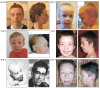
Craniofacial involvement. Typical facial characteristics (see Figure 1) can be present from early childhood, but usually become more prominent in older children and adults.

Generalized connective tissue disorder. The skin is usually soft and loose in ATS. Some affected individuals have a hyperextensible skin and rarely may present with frank cutis laxa [Callewaert et al 2008, Beyens et al 2018].

Individuals often present with hypotonia and joint hypermobility and are at risk for sprains and luxations. Adults are at increased risk for joint pain and fatigue [Castori et al 2012].

Diaphragmatic hernia and sliding hiatal hernias are reported in up to 50% of affected individuals [Callewaert et al 2008, Zaidi et al 2009].

Affected women are more prone to prolapse of the bladder, uterus, and rectum, especially following childbirth [Castori et al 2012].

Skeletal findings. Growth of the long bones may be excessive. Although clear dolichostenomelia (disproportionately long arms and legs compared to the trunk) is rarely present, overgrowth of the ribs may result in pectus deformity, and the hands often show arachnodactyly. Scoliosis is rare and ranges from mild to severe; it can be progressive, mostly during periods of fast growth. Pes planus with hindfoot valgus may be present. Knee and/or elbow contractures and camptodactyly have been reported.

Osteopenia has been observed in rare individuals [Authors, unpublished data].

Eye. Thin corneas and/or pellucid marginal degeneration of the corneas were present in five children assessed in detail [Hardin et al 2018]. Keratoconus has been reported in eight affected individuals, two of whom also had keratoglobus and deep corneal opacification [Callewaert et al 2008, Hasler et al 2011, Hardin et al 2018]. Note that the corneal findings become more pronounced with age, and that many children have not been assessed properly to identify corneal thinning (which often precedes keratoconus). Ectopia lentis has not been described. It is unclear whether myopia and astigmatism occur more frequently than in the general population.

Other

  • Respiratory tract. Infants may present with infant respiratory stress syndrome, requiring admission to a neonatal intensive care unit. Pulmonary hypertension may cause shortness of breath, fatigue, and cyanotic episodes. Single cases of early-onset emphysema [Takahashi et al 2013] and pneumonia [Wessels et al 2004] have been described, but it is unclear if individuals with ATS are truly at risk for these features.
  • Urogenital anomalies. Pyeloectasia and bladder diverticula have been reported in multiple individuals. Hypospadias, urine retention, and hematuria (underlying cause unknown) were each reported in single individuals.

Life span. Although early reports mentioned 40% mortality before age four years [Wessels et al 2004], larger series of individuals with a molecularly confirmed diagnosis indicate a milder disease spectrum [Callewaert et al 2008, Beyens et al 2018]. It is likely some individuals in whom the diagnosis was not molecularly confirmed had a similar disorder with a poorer prognosis – including EFEMP2-related cutis laxa (see Differential Diagnosis). The earlier literature may also have been biased toward reporting the more severe end of the phenotypic spectrum.

Genotype-Phenotype Correlations

No genotype-phenotype correlations have been identified.

Nomenclature

Lees et al [1969] were likely the first to report the syndrome, which they called "Ehlers-Danlos syndrome with multiple pulmonary artery stenoses and tortuous systemic arteries." Following this initial description, the disorder has always been considered distinct from Ehlers-Danlos syndrome, and the designation "arterial tortuosity syndrome" has been used consistently. Molecular genetic testing has not yet been performed in the individual published by Lees et al [1969].

Of note, in some reports "arterial tortuosity syndrome" refers to EFEMP2-related cutis laxa, which is a related but distinct disorder [Satish et al 2008].

Prevalence

No reliable estimates of prevalence exist. ATS is considered rare (<1:1,000,000 live births). However, some authors suggest that it may be more frequent than estimated [Callewaert et al 2008].

ATS occurs in all populations, but most reported cases to date are from Europe and the Middle East.

Differential Diagnosis

Table 3 summarizes disorders that should be considered in the differential diagnosis of arterial tortuosity syndrome (ATS).

Table 3.

Genes of Interest in the Differential Diagnosis of Arterial Tortuosity Syndrome

Gene(s)DiffDx DisorderMOIClinical Features of DiffDx DisorderDistinguishing Clinical Features
ACTA2
(BGN)
COL3A1
FBN1
(FOXE3)
(HCN4)
LOX
(MAT2A)
(MFAP5)
MYH11
MYLK
PRKG1
SMAD3
TGFB2
(TGFB3)
TGFBR1
TGFBR2 1
Heritable thoracic aortic disease (HTAD; familial thoracic aortic aneurysms) 1ADAortic root dilatation may be isolated or assoc w/other vascular & nonvascular features, incl marfanoid skeletal features as seen in ATS. 1Though some of these thoracic aortic aneurysm syndromes may present w/mild tortuosity, severe tortuosity is usually absent, & stenosis is rare (except in ACTA2-HTAD).
ALDH18A1 ALDH18A1 cutis laxa (ARCL3A: OMIM 219150; ADCL3: OMIM 616603)AR
AD 2
Intracerebral AT (no widespread AT); ID; chorea-athetosis; corneal clouding or cataract; intrauterine growth restriction; some degree of cutis laxaATS is not assoc w/ID, chorea-athetosis, corneal clouding, or cataract.
ATP7A Occipital horn syndrome (OHS) (See ATP7A Copper Transport Disorders.)XLOccipital horns 3 (may be clinically palpable or observed on skull radiographs); lax skin & joints; bladder diverticula; inguinal hernias; vascular tortuosity (mainly of cerebral vasculature)Skeletal & urogenital features of OHS are distinctive.
BGN BGN-associated aortic aneurysm syndrome 4XLClinical features significantly overlap w/Marfan syndrome & LDS: early-onset aortic root dilatation & dissection, widely spaced eyes, joint hypermobility, contractures, bifid uvula, & pectus deformities
  • Persons w/BGN-assoc aortic aneurysm syndrome may share craniofacial manifestations (widely spaced eyes, cleft uvula/palate, & craniosynostosis) w/persons w/LDS, findings less frequently seen in persons w/ATS.
  • AT is rarely present in BGN-assoc aortic aneurysm syndrome, and to a lesser degree than in ATS.
  • Persons w/BGN-assoc aortic aneurysm syndrome present more frequently w/aneurysm of aortic root than persons w/ATS.
COL3A1
TNXB
Hypermobile EDS (hEDS), TNXB-related classical-like EDS, & vascular EDS (vEDS)AD AR 5
  • hEDS: generalized joint hypermobility → repetitive joint luxations & chronic musculoskeletal pain; soft & hyperextensible skin; autonomic dysfunction
  • vEDS: thin, translucent skin; atrophic scars, easy bruising; characteristic facial appearance (in some persons); arterial, intestinal, &/or uterine fragility
  • Vascular dissection or rupture, GI perforation, or organ rupture are presenting signs in most adults w/vEDS.
  • AT is absent in persons w/hEDS & vEDS. 6
  • Skin & craniofacial features in vEDS are usually distinctive.
EFEMP2 EFEMP2 cutis laxa (ARCL1B)AR
  • Cutis laxa & systemic involvement, most commonly AT, aneurysms, & stenosis; retrognathia; joint laxity; arachnodactyly
  • Severity ranges from perinatal lethality (due to cardiopulmonary failure) to manifestations limited to vascular & craniofacial systems
  • Focal stenosis at aortic isthmus is more common in ARCL1B than in ATS. 7
  • ARCL1B often presents w/more aggressive arterial phenotype w/rapid progression to aneurysms.
  • Typical facial characteristics of ATS (e.g., blepharophimosis, convex nasal ridge, & long face) are often absent in ARCL1B.
EMILIN1 EMILIN1-related cutis laxa 8Cutis laxa, AT, aortic root aneurysms
  • Osteopenia w/neonatal fractures
  • Typical facial characteristics of ATS are absent
FBLN5
LTBP4
FBLN5 cutis laxa & LTBP4 cutis laxaAR
AD 9
  • Cutis laxa, early childhood-onset pulmonary emphysema, peripheral pulmonary artery stenosis, & other evidence of generalized connective disorder incl inguinal hernias & hollow viscus diverticula (e.g., intestine, bladder)
  • Supravalvar aortic stenosis occasionally observed
  • Pulmonary emphysema or GI ruptures are often cause of death.
Persons w/FBLN5 cutis laxa & LTBP4 cutis laxa do not have the AT seen in persons w/ATS. 10
FBN1 Marfan syndrome ADAortic root aneurysm; long bone overgrowth; scoliosis; lens subluxation
  • Persons w/Marfan syndrome do not show manifest AT.
  • Lens subluxation is not present in ATS.
FKBP1
PLOD1
Kyphoscoliotic EDS (See FKBP14-kEDS & PLOD1-kEDS.)ARAortic root aneurysm; vascular rupture; scoliosis; corneal thinning; ocular rupture; joint hypermobility; skin fragility
  • Persons w/kEDS do not present w/manifest tortuosity.
  • Ocular fragility (a feature of PLOD1-kEDS) is not seen in ATS.
SMAD2
SMAD3
TGFB2
TGFB3
TGFBR1
TGFBR2
Loeys-Dietz syndrome (LDS)AD
  • Vascular findings (cerebral, thoracic, & abdominal arterial aneurysms, &/or dissections)
  • Skeletal manifestations (pectus excavatum or pectus carinatum, scoliosis, joint laxity, arachnodactyly, talipes equinovarus)
  • Persons w/LDS may have craniofacial manifestations (widely spaced eyes, cleft uvula/palate, craniosynostosis), findings usually not seen w/ATS.
  • AT is often present in LDS but more often in ATS.
  • Persons w/LDS present more frequently w/aneurysm of aortic root than persons w/ATS.

AD = autosomal dominant; AR = autosomal recessive; AT = arterial tortuosity; ATS = arterial tortuosity syndrome; DiffDx = differential diagnosis; EDS = Ehlers-Danlos syndrome; GI = gastrointestinal; ID = intellectual disability; LDS = Loeys-Dietz syndrome; MOI = mode of inheritance; XL = X-linked

1.

Renard et al [2018] discussed the clinical validity of genes associated with heritable thoracic aortic aneurysm and dissection (FTAAD). Genes in ( )s were recently associated with FTAAD and require further confirmation. Of note, this paper mentions SLC2A10 as a "potentially diagnostic gene for FTAAD," meaning that identification of biallelic SLC2A10 pathogenic variants may allow diagnosis of the cause of thoracic aortic enlargement, but that pathogenic variants in SLC2A10 are primarily causative of other clinical features and are not associated with a significant risk of progression to aortic dissection.

2.

ALDH18A1-related cutis laxa can be inherited in an autosomal recessive or autosomal dominant manner; the autosomal recessive form is more severe.

3.

"Occipital horns" are distinctive wedge-shaped calcifications at the sites of attachment of the trapezius muscle and the sternocleidomastoid muscle to the occipital bone.

4.
5.

Hypermobile EDS is inherited in an autosomal dominant manner (the molecular basis of hEDS is unknown). TNXB-related classical-like EDS is inherited in an autosomal recessive manner. Vascular EDS is associated with pathogenic variants in COL3A1 and is almost always inherited in an autosomal dominant manner (rare examples of biallelic inheritance have been reported).

6.

Two reports mention arterial tortuosity syndrome in individuals previously misdiagnosed with EDS hypermobility type, one of whom was homozygous for the SLC2A10 pathogenic variant c.1411+1G>A [Allen et al 2009] and one of whom was homozygous for the SLC2A10 pathogenic variant c.685C>T [Castori et al 2012].

7.
8.
9.

LTBP4-related cutis laxa is inherited in an autosomal recessive manner. FBLN5 cutis laxa can be inherited in an autosomal recessive or (less commonly) autosomal dominant manner.

10.

Management

No clinical practice guidelines for arterial tortuosity syndrome (ATS) have been published.

Evaluations Following Initial Diagnosis

To establish the extent of disease and needs in an individual diagnosed with ATS, the evaluations summarized in Table 4 (if not performed as part of the evaluation that led to the diagnosis) are recommended.

Table 4.

Recommended Evaluations Following Initial Diagnosis in Individuals with Arterial Tortuosity Syndrome

System/ConcernEvaluationComment
Aortic root aneurysm EchocardiographyAortic root measurements must be interpreted based on consideration of normal values for age & body size.
Arterial tortuosity (AT) MRA or CT scan w/3D reconstruction from head to pelvisTo evaluate AT, identify abnormal implantation of aortic branches & identify arterial aneurysms &/or stenosis throughout arterial tree.
Hypertension Blood pressure measurements
Orthostatic hypotension Tilt testingOnly if history is suggestive
Palate abnormality Eval for cleft palate or bifid uvula
Diaphragmatic hernia /
Sliding hernia
Thoracoabdominal radiography
Scoliosis Clinical eval, skeletal radiographs
Osteopenia Bone densitometryIf diagnosed in adulthood
Ocular abnormality Eval for keratoconus, keratoglobus & corneal thinning, refractive errors
Urogenital abnormalities Ultrasound of urinary tract
Genetic counseling By genetics professionals 1To inform affected persons & their families re nature, MOI, & implications of ATS to facilitate medical & personal decision making

ATS = arterial tortuosity syndrome; MOI = mode of inheritance

1.

Medical geneticist, certified genetic counselor, or certified advanced genetic nurse

Treatment of Manifestations

Individuals with ATS benefit from a coordinated approach from multidisciplinary specialists including a clinical geneticist, cardiologist, ophthalmologist, orthopedist, and cardiothoracic surgeon. If feasible, individuals with ATS should be managed in a medical center familiar with this condition.

Table 5.

Treatment of Manifestations in Individuals with Arterial Tortuosity Syndrome

Manifestation/
Concern
TreatmentConsiderations/Other
Aortic
aneurysm
Medical treatment: beta-adrenergic blockers or other medications, incl angiotensin-converting enzyme inhibitors (ACE-I) & angiotensin II receptor 1 (ATIIR1) antagonists such as losartan
  • Efficacy of these treatments has not been established in ATS.
  • Caution is warranted when using blood pressure-lowering medications in presence of arterial stenosis (anatomic or functional due to severe tortuosity), esp renal artery stenosis, as such meds may confer risk for renal failure.
Surgical treatment: valve-sparing procedure that precludes need for chronic anticoagulation
  • No data available on aortic diameter at which intervention is appropriate
  • Decision making should also incl assessment of family history or individual's personal assessment of risk vs benefit.
Focal stenosis
of aorta & aortic
branches
Catheterization &/or surgery
Pulmonary artery
stenosis →
pulmonary
hypertension
Surgery, catheterization, or a hybrid (transcatheter-surgical procedure)
Abnormal
skin healing
Careful postoperative eval of (surgical) wounds needed, as wound healing may be delayedStitches should avoid traction & should remain in place for ~10 days.
Hernias Use of supporting mesh during surgical repair to minimize recurrence
Pectus
excavatum
Surgical intervention when severeSurgery would be medically (rather than cosmetically) indicated.
Scoliosis Mgmt by orthopedistSurgery may be needed.
Pes planus Orthotics
Ocular issues Mgmt by ophthalmologist w/expertise in connective tissue disordersTo incl aggressive refraction & visual correction & eval of keratoconus
Emphysema Treat symptomaticallyPositive pressure ventilation may cause emphysematous changes to progress.
General
well-being
  • Persons can & should maintain aerobic activity (e.g., swimming) in moderation.
  • Persons w/ATS may have psychological issues, esp in adolescence, for which psychological guidance may be relevant.

ATS = arterial tortuosity syndrome

Surveillance

Table 6.

Recommended Surveillance for Individuals with Arterial Tortuosity Syndrome

System/ConcernEvaluationFrequency
Arterial
tortuosity,
aneurysms,
stenosis, or
dilatation of
aortic root
Echocardiography
  • Every 3 mos until age 5 yrs
  • Annually thereafter if diameters of aorta are w/in normal limits
MRA or CT scan w/3D reconstructions from head to pelvis
  • Annually from birth or time of diagnosis
  • Under stable conditions (in absence of aneurysms, stenosis, or dilatation of aortic root), perform every 3 yrs in older children & adults.
Pulmonary
hypertension
EchocardiogramPer cardiologist
Systemic
hypertension
Blood pressure measurementAt each visit
Dental
crowding
Orthodontic evalDuring eruption of permanent dentition
Scoliosis Radiographs to evaluate for progression of scoliosisDuring periods of rapid growth (first 2 yrs of life & during puberty)
Ocular issues Routine follow up for refractive errors & keratoconus w/ophthalmologist w/expertise in connective tissue disorders
Emphysema Eval w/pulmonologist
  • Baseline lung function at age 18 yrs
  • Repeat exam based on symptomatology

Agents/Circumstances to Avoid

Avoid the following:

  • Contact sports, competitive sports, and isometric exercise
  • Scuba diving because of pressure differences and the need for positive pressure ventilation
  • Agents that stimulate the cardiovascular system, including routine use of decongestants
  • Use of tobacco, which increases cardiovascular and pulmonary risk and the likelihood of premature skin aging
  • Sun tanning, which increases the likelihood of premature skin aging

Evaluation of Relatives at Risk

It is appropriate to evaluate the older and younger sibs of a proband with ATS in order to identify as early as possible those who would benefit from institution of surveillance and preventive measures.

  • If the SLC2A10 pathogenic variants in the family are known, molecular genetic testing can be used to clarify the genetic status of at-risk sibs.
  • If the pathogenic variants in the family are not known, at-risk sibs should be evaluated for signs of the disorder (clinically and with echocardiography or more advanced vascular imaging, as clinical symptoms may be very subtle) to clarify their genetic status.

See Genetic Counseling for issues related to testing of at-risk relatives for genetic counseling purposes.

Pregnancy Management

Data on the management of women with arterial tortuosity syndrome during pregnancy and delivery are limited, with only three pregnancies reported (in 2 women) to date. All pregnancies had a good outcome; however, one woman experienced pelvic organ prolapse following the vaginal delivery of her first child [Castori et al 2012]. The other woman, who delivered without complications at 34 weeks' gestation by elective cæsarean section, was started on acetylsalicylic acid to prevent thromboembolic events eight days post partum [Allen et al 2009].

Preconception counseling should include possible pregnancy-associated risks to the mother and medication-associated risks to the fetus.

The risks to the mother are mainly those of aortic root dilatation and dissection. As is common practice in management in Marfan syndrome, elective aortic repair using a valve-sparing procedure (if possible) could be performed prior to conception when the aortic root diameter reaches 45 mm.

Currently, no data are available on a possible risk for pregnancy-associated uterine rupture (as is seen in Loeys-Dietz syndrome and EDS, vascular type). Prenatal and postnatal physiotherapy can minimize the risk for pelvic organ prolapse.

Peripartum intensive monitoring is advised. Pregnancies should be followed by a high-risk obstetrician and a cardiologist familiar with this or related conditions.

Increased surveillance of the aortic root and previously detected aneurysms during pregnancy and following delivery is recommended because of the increased risk for progressive dilatation. Echocardiography is suggested every two to three months from conception until six months post partum.

Delivery should be planned in a center with experience with this or related conditions. It is currently unclear whether cæsarean section or vaginal delivery is preferable.

Medication-associated risks to the fetus. The effects of angiotensin-converting enzyme inhibitors (ACE-I) on the fetus in the first trimester of pregnancy are incompletely understood; however, use in the second and third trimesters of pregnancy can lead to fetal death and damage. Angiotensin II receptor 1 antagonists (ATIIR1) such as losartan are thought to cause fetal effects similar to ACE-I (including fetal damage, oligohydramnios, and fetal death) if taken during the second and/or third trimesters of pregnancy.

  • Ideally, women with ATS who are planning a pregnancy should transition to a different antihypertensive medication (e.g., a beta-blocker) prior to conception.
  • Women with ATS who become pregnant while taking an ACE-I or an ATIIR1 should be transitioned to a different antihypertensive medication as soon as the pregnancy is recognized.

Women undergoing a non-valve-sparing aortic root replacement before pregnancy should be advised of the risk associated with anticoagulant therapy during pregnancy.

See MotherToBaby for further information on medication use during pregnancy.

Therapies Under Investigation

Search ClinicalTrials.gov in the US and EU Clinical Trials Register in Europe for access to information on clinical studies for a wide range of diseases and conditions. Note: There may not be clinical trials for this disorder.

Genetic Counseling

Genetic counseling is the process of providing individuals and families with information on the nature, mode(s) of inheritance, and implications of genetic disorders to help them make informed medical and personal decisions. The following section deals with genetic risk assessment and the use of family history and genetic testing to clarify genetic status for family members; it is not meant to address all personal, cultural, or ethical issues that may arise or to substitute for consultation with a genetics professional. —ED.

Mode of Inheritance

Arterial tortuosity syndrome (ATS) is inherited in an autosomal recessive manner.

Risk to Family Members

Parents of a proband

  • The parents of an affected individual are obligate heterozygotes (i.e., presumed to be carriers of one SLC2A10 pathogenic variant based on family history).
  • Molecular genetic testing is recommended for the parents of a proband to confirm that both parents are heterozygous for an SLC2A10 pathogenic variant and to allow reliable recurrence risk assessment. (In rare families, only one parent is heterozygous and the proband is affected as the result of either: (1) one pathogenic variant inherited from the heterozygous parent and a second pathogenic variant that occurred de novo in the proband; or (2) uniparental isodisomy and consequent homozygosity for the pathogenic variant transmitted by a heterozygous parent [Robinson 2000, Jónsson et al 2017].)
  • Heterozygotes (carriers) are asymptomatic and are not at risk of developing the disorder.

Sibs of a proband

  • If both parents are known to be heterozygous for an SLC2A10 pathogenic variant, each sib of an affected individual has at conception a 25% chance of being affected, a 50% chance of being an asymptomatic carrier, and a 25% chance of being unaffected and not a carrier.
  • Heterozygotes (carriers) are asymptomatic and are not at risk of developing arterial tortuosity syndrome.

Offspring of a proband. The offspring of an individual with ATS are obligate heterozygotes (carriers) for a pathogenic variant in SLC2A10.

Other family members. Each sib of the proband's parents is at a 50% risk of being a carrier of an SLC2A10 pathogenic variant.

Carrier Detection

Carrier testing for at-risk relatives requires prior identification of the SLC2A10 pathogenic variants in the family.

Related Genetic Counseling Issues

See Management, Evaluation of Relatives at Risk for information on evaluating at-risk relatives for the purpose of early diagnosis and treatment.

Family planning

  • The optimal time for determination of genetic risk, clarification of carrier status, and discussion of the availability of prenatal/preimplantation genetic testing is before pregnancy.
  • It is appropriate to offer genetic counseling (including discussion of potential risks to offspring and reproductive options) to young adults who are affected, are carriers, or are at risk of being carriers.

DNA banking. Because it is likely that testing methodology and our understanding of genes, pathogenic mechanisms, and diseases will improve in the future, consideration should be given to banking DNA from probands in whom a molecular diagnosis has not been confirmed (i.e., the causative pathogenic mechanism is unknown). For more information, see Huang et al [2022].

Prenatal Testing and Preimplantation Genetic Testing

Once the SLC2A10 pathogenic variants have been identified in an affected family member, prenatal and preimplantation genetic testing for ATS are possible.

Differences in perspective may exist among medical professionals and within families regarding the use of prenatal testing. While most centers would consider use of prenatal testing to be a personal decision, discussion of these issues may be helpful.

Resources

GeneReviews staff has selected the following disease-specific and/or umbrella support organizations and/or registries for the benefit of individuals with this disorder and their families. GeneReviews is not responsible for the information provided by other organizations. For information on selection criteria, click here.

Molecular Genetics

Information in the Molecular Genetics and OMIM tables may differ from that elsewhere in the GeneReview: tables may contain more recent information. —ED.

Table A.

Arterial Tortuosity Syndrome: Genes and Databases

Data are compiled from the following standard references: gene from HGNC; chromosome locus from OMIM; protein from UniProt. For a description of databases (Locus Specific, HGMD, ClinVar) to which links are provided, click here.

Table B.

OMIM Entries for Arterial Tortuosity Syndrome (View All in OMIM)

208050ARTERIAL TORTUOSITY SYNDROME; ATORS
606145SOLUTE CARRIER FAMILY 2 (FACILITATED GLUCOSE TRANSPORTER), MEMBER 10; SLC2A10

Molecular Pathogenesis

SLC2A10 encodes for the facilitative glucose transporter, GLUT10. Over the last several years, accumulating evidence has identified GLUT10 as an intracellular transporter of dehydroascorbic acid (DHA). In the endoplasmic reticulum, DHA is required as a cofactor for prolyl and lysyl hydroxylases for the crosslinking of collagen and elastin monomers. In the mitochondria, DHA aids in maintaining redox homeostasis and scavenging reactive oxygen species. Finally, in the nucleoplasm, ascorbate activates Fe2+/2-oxoglutarate-dependent dioxygenases (which require ascorbate as a cofactor) that participate in epigenetic regulation of gene expression via histone and DNA demethylation. Hence, arterial tortuosity syndrome emerges as an ascorbate compartmentalization disorder in which the pathogenesis may affect many physiologic processes in different cellular organelles. For a comprehensive overview, see Boel et al [2021].

Mechanism of disease causation. Arterial tortuosity syndrome is caused by loss-of-function variants in SLC2A10 that lead to a loss of function of the protein GLUT10.

Chapter Notes

Author Notes

University of Ghent Center for Medical Genetics

Bert Callewaert is an assistant professor at Ghent University, and pediatrician and clinical geneticist at the Center for Medical Genetics, Ghent University Hospital. He is a senior clinical investigator supported by the Research Foundation – Flanders. His research focuses on clinical aspects and mechanisms of disease in connective tissue disorders including the arterial tortuosity syndrome, cutis laxa syndromes, congenital contractural arachnodactyly, and familial thoracic aortic aneurysms.

Paul Coucke is the supervisor of the connective tissue laboratory and head of the department at the Center for Medical Genetics.

Anne De Paepe is a clinical geneticist at the Center for Medical Genetics.

Dr Callewaert (bert.callewaert@ugent.be) is actively involved in clinical research regarding individuals with arterial tortuosity syndrome. He would be happy to communicate with persons who have any questions regarding diagnosis of arterial tortuosity syndrome or other considerations.

Contact Dr Callewaert at bert.callewaert@ugent.be to inquire about the interpretation of SLC2A10 variants of uncertain significance.

Dr Callewaert (bert.callewaert@ugent.be) is also interested in hearing from clinicians treating families affected by arterial tortuosity syndrome in whom no causative variant has been identified through molecular genetic testing.

Acknowledgments

Bert Callewaert is a senior clinical investigator of the Fund for Scientific Research-Flanders.

Revision History

References

Literature Cited

  • Adamo CS, Beyens A, Schiavinato A, Keene DR, Tufa SF, Mörgelin M, Brinckmann J, Sasaki T, Niehoff A, Dreiner M, Pottie L, Muiño-Mosquera L, Gulec EY, Gezdirici A, Braghetta P, Bonaldo P, Wagener R, Paulsson M, Bornaun H, De Rycke R, De Bruyne M, Baeke F, Devine WP, Gangaram B, Tam A, Balasubramanian M, Ellard S, Moore S, Symoens S, Shen J, Cole S, Schwarze U, Holmes KW, Hayflick SJ, Wiszniewski W, Nampoothiri S, Davis EC, Sakai LY, Sengle G, Callewaert B. EMILIN1 deficiency causes arterial tortuosity with osteopenia and connects impaired elastogenesis with defective collagen fibrillogenesis. Am J Hum Genet. 2022;109:2230–52. [PMC free article: PMC9748297] [PubMed: 36351433]
  • Allen VM, Horne SG, Penney LS, Rapchuk IL, Brock JA, Thompson DL, Stinson DA. Successful outcome in pregnancy with arterial tortuosity syndrome. Obstet Gynecol. 2009;114:494–8. [PubMed: 19622975]
  • Beyens A, Albuisson J, Boel A, Al-Essa M, Al-Manea W, Bonnet D, Bostan O, Boute O, Busa T, Canham N, Cil E, Coucke PJ, Cousin MA, Dasouki M, De Backer J, De Paepe A, De Schepper S, De Silva D, Devriendt K, De Wandele I, Deyle DR, Dietz H, Dupuis-Girod S, Fontenot E, Fischer-Zirnsak B, Gezdirici A, Ghoumid J, Giuliano F, Diéz NB, Haider MZ, Hardin JS, Jeunemaitre X, Klee EW, Kornak U, Landecho MF, Legrand A, Loeys B, Lyonnet S, Michael H, Moceri P, Mohammed S, Muiño-Mosquera L, Nampoothiri S, Pichler K, Prescott K, Rajeb A, Ramos-Arroyo M, Rossi M, Salih M, Seidahmed MZ, Schaefer E, Steichen-Gersdorf E, Temel S, Uysal F, Vanhomwegen M, Van Laer L, Van Maldergem L, Warner D, Willaert A, Collins TR, Taylor A, Davis EC, Zarate Y, Callewaert B. Arterial tortuosity syndrome: 40 new families and literature review. Genet Med. 2018;20:1236–45. [PubMed: 29323665]
  • Boel A, Veszelyi K, Németh CE, Beyens A, Willaert A, Coucke P, Callewaert B, Margittai É. Arterial tortuosity syndrome: an ascorbate compartmentalization disorder? Antioxid Redox Signal. 2021;34:875–89. [PubMed: 31621376]
  • Bottio T, Bisleri G, Piccoli P, Muneretto C. Valve-sparing aortic root replacement in a patient with a rare connective tissue disorder: arterial tortuosity syndrome. J Thorac Cardiovasc Surg. 2007;133:252–3. [PubMed: 17198824]
  • Callewaert B, Su CT, Van Damme T, Vlummens P, Malfait F, Vanakker O, Schulz B, Mac Neal M, Davis EC, Lee JG, Salhi A, Unger S, Heimdal K, De Almeida S, Kornak U, Gaspar H, Bresson JL, Prescott K, Gosendi ME, Mansour S, Piérard GE, Madan-Khetarpal S, Sciurba FC, Symoens S, Coucke PJ, Van Maldergem L, Urban Z, De Paepe A. Comprehensive clinical and molecular analysis of 12 families with type 1 recessive cutis laxa. Hum Mutat. 2013;34:111–21. [PMC free article: PMC4105850] [PubMed: 22829427]
  • Callewaert BL, Willaert A, Kerstjens-Frederikse WS, De Backer J, Devriendt K, Albrecht B, Ramos-Arroyo MA, Doco-Fenzy M, Hennekam RC, Pyeritz RE, Krogmann ON, Gillessen-kaesbach G, Wakeling EL, Nik-zainal S, Francannet C, Mauran P, Booth C, Barrow M, Dekens R, Loeys BL, Coucke PJ, De Paepe AM. Arterial tortuosity syndrome: clinical and molecular findings in 12 newly identified families. Hum Mutat. 2008;29:150–8. [PubMed: 17935213]
  • Castori M, Ritelli M, Zoppi N, Molisso L, Chiarelli N, Zaccagna F, Grammatico P, Colombi M. Adult presentation of arterial tortuosity syndrome in a 51-year-old woman with a novel homozygous c.1411+1G>A mutation in the SLC2A10 gene. Am J Med Genet A. 2012;158A:1164–9. [PubMed: 22488877]
  • Coucke PJ, Willaert A, Wessels MW, Callewaert B, Zoppi N, De Backer J, Fox JE, Mancini GM, Kambouris M, Gardella R, Facchetti F, Willems PJ, Forsyth R, Dietz HC, Barlati S, Colombi M, Loeys B, De Paepe A. Mutations in the facilitative glucose transporter GLUT10 alter angiogenesis and cause arterial tortuosity syndrome. Nat Genet. 2006;38:452–7. [PubMed: 16550171]
  • de Marcellus C, Baudouin V, Tanase A, Monet C, Perrin L, Deschenes G, Hogan J. Severe neonatal hypertension revealing arterial tortuosity syndrome. Kidney Int. 2018;93:526. [PubMed: 29389400]
  • Drera B, Barlati S, Colombi M. Ischemic stroke in an adolescent with arterial tortuosity syndrome. Neurology. 2007a;68:1637. [PubMed: 17485657]
  • Drera B, Guala A, Zoppi N, Gardella R, Franceschini P, Barlati S, Colombi M. Two novel SLC2A10/GLUT10 mutations in a patient with arterial tortuosity syndrome. Am J Med Genet A. 2007b;143A:216–8. [PubMed: 17163528]
  • Hardin JS, Zarate YA, Callewaert B, Phillips PH, Warner DB. Ophthalmic findings in patients with arterial tortuosity syndrome and carriers: a case series. Ophthalmic Genet. 2018;39:29–34. [PubMed: 28726533]
  • Hasler S, Stürmer J, Kaufmann C. Keratoglobus and deep stromal corneal opacification in a case of arterial tortuosity syndrome. Klin Monbl Augenheilkd. 2011;228:345–6. [PubMed: 21484644]
  • Huang SJ, Amendola LM, Sternen DL. Variation among DNA banking consent forms: points for clinicians to bank on. J Community Genet. 2022;13:389–97. [PMC free article: PMC9314484] [PubMed: 35834113]
  • Hucthagowder V, Sausgruber N, Kim KH, Angle B, Marmorstein LY, Urban Z. Fibulin-4: a novel gene for an autosomal recessive cutis laxa syndrome. Am J Hum Genet. 2006;78:1075–80. [PMC free article: PMC1474103] [PubMed: 16685658]
  • Jónsson H, Sulem P, Kehr B, Kristmundsdottir S, Zink F, Hjartarson E, Hardarson MT, Hjorleifsson KE, Eggertsson HP, Gudjonsson SA, Ward LD, Arnadottir GA, Helgason EA, Helgason H, Gylfason A, Jonasdottir A, Jonasdottir A, Rafnar T, Frigge M, Stacey SN, Th Magnusson O, Thorsteinsdottir U, Masson G, Kong A, Halldorsson BV, Helgason A, Gudbjartsson DF, Stefansson K. Parental influence on human germline de novo mutations in 1,548 trios from Iceland. Nature. 2017;549:519–22. [PubMed: 28959963]
  • Kocova M, Kacarska R, Kuzevska-Maneva K, Prijic S, Lazareska M, Dordoni C, Ritelli M, Colombi M. Clinical variability in two Macedonian families with arterial tortuosity syndrome. Balkan J Med Genet. 2018;21:47–52. [PMC free article: PMC6231313] [PubMed: 30425910]
  • Lees MH, Menashe VD, Sunderland CO, Mgan CL, Dawson PJ. Ehlers-Danlos syndrome associated with multiple pulmonary artery stenoses and tortuous systemic arteries. J Pediatr. 1969;75:1031–6. [PubMed: 5352829]
  • Meester JA, Vandeweyer G, Pintelon I, Lammens M, Van Hoorick L, De Belder S, Waitzman K, Young L, Markham LW, Vogt J, Richer J, Beauchesne LM, Unger S, Superti-Furga A, Prsa M, Dhillon R, Reyniers E, Dietz HC, Wuyts W, Mortier G, Verstraeten A, Van Laer L, Loeys BL. Loss-of-function mutations in the X-linked biglycan gene cause a severe syndromic form of thoracic aortic aneurysms and dissections. Genet Med. 2017;19:386–95. [PMC free article: PMC5207316] [PubMed: 27632686]
  • Moceri P, Albuisson J, Saint-Faust M, Casagrande F, Giuliano F, Devos C, Benoit P, Hugues N, Ducreux D, Cerboni P, Dageville C, Jeunemaitre X. Arterial tortuosity syndrome: early diagnosis and association with venous tortuosity. J Am Coll Cardiol. 2013;61:783. [PubMed: 23410549]
  • Pletcher BA, Fox JE, Boxer RA, Singh S, Blumenthal D, Cohen T, Brunson S, Tafreshi P, Kahn E. Four sibs with arterial tortuosity: description and review of the literature. Am J Med Genet. 1996;66:121–8. [PubMed: 8958317]
  • Renard M, Francis C, Ghosh R, Scott AF, Witmer PD, Adès LC, Andelfinger GU, Arnaud P, Boileau C, Callewaert BL, Guo D, Hanna N, Lindsay ME, Morisaki H, Morisaki T, Pachter N, Robert L, Van Laer L, Dietz HC, Loeys BL, Milewicz DM, De Backer J. Clinical validity of genes for heritable thoracic aortic aneurysm and dissection. J Am Coll Cardiol. 2018;72:605–15. [PMC free article: PMC6378369] [PubMed: 30071989]
  • Renard M, Holm T, Veith R, Callewaert BL, Adès LC, Baspinar O, Pickart A, Dasouki M, Hoyer J, Rauch A, Trapane P, Earing MG, Coucke PJ, Sakai LY, Dietz HC, De Paepe AM, Loeys BL. Altered TGFbeta signaling and cardiovascular manifestations in patients with autosomal recessive cutis laxa type I caused by fibulin-4 deficiency. Eur J Hum Genet. 2010;18:895–901. [PMC free article: PMC2987390] [PubMed: 20389311]
  • Richards S, Aziz N, Bale S, Bick D, Das S, Gastier-Foster J, Grody WW, Hegde M, Lyon E, Spector E, Voelkerding K, Rehm HL, et al. Standards and guidelines for the interpretation of sequence variants: a joint consensus recommendation of the American College of Medical Genetics and Genomics and the Association for Molecular Pathology. Genet Med. 2015;17:405–24. [PMC free article: PMC4544753] [PubMed: 25741868]
  • Robinson WP. Mechanisms leading to uniparental disomy and their clinical consequences. Bioessays. 2000;22:452–9. [PubMed: 10797485]
  • Satish G, Nampoothiri S, Kappanayil M. Images in cardiovascular medicine. Arterial tortuosity syndrome: phenotypic features and cardiovascular manifestations. Circulation. 2008;117:e477–8. [PubMed: 18541745]
  • Stenson PD, Mort M, Ball EV, Chapman M, Evans K, Azevedo L, Hayden M, Heywood S, Millar DS, Phillips AD, Cooper DN. The Human Gene Mutation Database (HGMD®): optimizing its use in a clinical diagnostic or research setting. Hum Genet. 2020;139:1197–207. [PMC free article: PMC7497289] [PubMed: 32596782]
  • Takahashi Y, Fujii K, Yoshida A, Morisaki H, Kohno Y, Morisaki T. Artery tortuosity syndrome exhibiting early-onset emphysema with novel compound heterozygous SLC2A10 mutations. Am J Med Genet A. 2013;161A:856–9. [PubMed: 23494979]
  • Wessels MW, Catsman-Berrevoets CE, Mancini GM, Breuning MH, Hoogeboom JJ, Stroink H, Frohn-Mulder I, Coucke PJ, Paepe AD, Niermeijer MF, Willems PJ. Three new families with arterial tortuosity syndrome. Am J Med Genet A. 2004;131:134–43. [PubMed: 15529317]
  • Zaidi SH, Meyer S, Peltekova I, Teebi AS, Faiyaz-Ul-Haque M. Congenital diaphragmatic abnormalities in arterial tortuosity syndrome patients who carry mutations in the SLC2A10 gene. Clin Genet. 2009;75:588–9. [PubMed: 19508422]
  • Zoma JM, Aggarwal S, Kobayashi D. Arterial tortuosity syndrome in a term neonate. Ann Thorac Surg. 2019;108:e281. [PubMed: 31077662]
Copyright © 1993-2024, University of Washington, Seattle. GeneReviews is a registered trademark of the University of Washington, Seattle. All rights reserved.

GeneReviews® chapters are owned by the University of Washington. Permission is hereby granted to reproduce, distribute, and translate copies of content materials for noncommercial research purposes only, provided that (i) credit for source (http://www.genereviews.org/) and copyright (© 1993-2024 University of Washington) are included with each copy; (ii) a link to the original material is provided whenever the material is published elsewhere on the Web; and (iii) reproducers, distributors, and/or translators comply with the GeneReviews® Copyright Notice and Usage Disclaimer. No further modifications are allowed. For clarity, excerpts of GeneReviews chapters for use in lab reports and clinic notes are a permitted use.

For more information, see the GeneReviews® Copyright Notice and Usage Disclaimer.

For questions regarding permissions or whether a specified use is allowed, contact: ude.wu@tssamda.

Bookshelf ID: NBK253404PMID: 25392904

Views

Tests in GTR by Gene

Related information

  • OMIM
    Related OMIM records
  • PMC
    PubMed Central citations
  • PubMed
    Links to PubMed
  • Gene
    Locus Links

Similar articles in PubMed

See reviews...See all...

Recent Activity

Your browsing activity is empty.

Activity recording is turned off.

Turn recording back on

See more...