

NCBI Bookshelf. A service of the National Library of Medicine, National Institutes of Health.
Dean L, McEntyre J, editors. Coffee Break: Tutorials for NCBI Tools [Internet]. Bethesda (MD): National Center for Biotechnology Information (US); 1999-.

As humans age, many bodily functions and abilities change. Scientists have long been interested in discovering how aging produces a decline in brain function and contributes to the development of certain diseases. To investigate the molecular shifts that occur in the brain during aging, some recent studies have focused on changes in gene expression patterns, both in humans and in other animals.
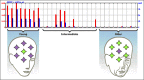
A recent study by Lu et al. (1) compared the levels of gene expression in postmortem brain samples from young and old human subjects. The gene expression profiles were clustered into three broad groups: young (below 40 years), intermediate (40-70 years), and old (70+ years) (see data at the Gene Expression Omnibus) (2). Whereas the young and old subjects were relatively homogeneous in their gene expression profiles, the intermediate group was much more heterogeneous, with vastly different rates of progression through the transition from the young gene expression profile to the old profile.
In older brains, the expression of genes involved in learning and memory, neuronal survival, and maintenance was decreased. Among these were the genes coding for microtubule-associated protein MAP1B, which stabilizes microtubules and promotes axonal growth (3,4) (see Entrez Gene entry, GEO data); MEF2C (see Entrez Gene entry, GEO data), which promotes the survival of neurons (1); subunit 2A of the glutamate receptor NMDAR (see Entrez Gene entry, GEO data), which is centrally involved in synaptic plasticity, the brain-restructuring process used in learning and memory; and calmodulin 1 (see Entrez Gene entry, GEO data), which is a central regulator of calcium-mediated signaling and plays an important role in memory. In contrast, the expression of genes coding for proteins involved in sensing and responding to cellular stresses increased, suggesting that older brains are exposed to higher levels of damaging stressors than are young brains (1). For example, one of the genes more highly expressed in older brains was the DNA repair enzyme OGG1 (see Entrez Gene entry, GEO data), which targets oxidatively damaged DNA.
Similar studies have been performed with a range of other animals, including Caenorhabditis elegans, Drosophila melogaster (5), and mice (6,7). Although the affected genes were not the same among all of the systems, the functional systems of the cells were affected in similar ways across species. For example, in the brains of both mice and humans, the expression of genes involved in synaptic function was decreased, whereas those involved in stress responses increased.
Further investigation of the genes whose expression was decreased in older human subjects showed that the promoters of these genes were more susceptible to oxidative damage than other genes tested (1), suggesting a mechanism for the observed decreased gene expression. Oxidative damage is caused by oxidants such as superoxide and hydrogen peroxide, which are produced as natural by-products of cell metabolism (8). The cell has response systems specifically dedicated to sensing and destroying such oxidants, as well as systems to repair any damage caused by those oxidants. It has long been hypothesized that oxidants play a role in aging processes, but this has proved difficult to demonstrate directly (9). The variable gene expression profiles (1) of the intermediate-age group suggest that oxidative damage may accumulate over a long period of time, with the effects occurring long after the initial oxidative damage is inflicted.
Perhaps some of the effects of aging can be slowed or lessened by controlling the level of oxidative stress in the cell; many methods are currently under investigation for reducing the creation of oxidants during metabolism. These include calorie restriction (currently being tested in clinical trials, see news coverage) (10), the use of antioxidant supplements, and a wide range of methods designed to target various cellular and molecular processes to safely and effectively reduce the production of oxidants during metabolism (10). Another potentially fruitful therapeutic strategy is to enhance DNA repair in cells to slow the accumulation of oxidative damage in the DNA. Because the results outlined by Lu et al. (10) show that after the age of about 40 years, humans may begin to exhibit age-related changes in gene expression, some treatments might be more effective in young patients than in those already experiencing the effects of aging. The challenge for future scientific investigation will be to find effective treatments for increasing life expectancy while ensuring high quality of life.
This Coffee Break was contributed by Victoria Sutton, PhD, while on rotation at the National Center for Biotechnology Information as a part of the Emerging Leaders Program from the Department of Health and Human Services (DHSS).
Aging and the Human Brain
The changes in gene expression that underlie the effects of aging in humans
For this tutorial, you will need the latest version of Flash player installed on your computer.
References
- 1.
- Lu T . et al. Gene regulation and DNA damage in the ageing human brain. Nature. 2004;429:883–891. [PubMed: 15190254]
- 2.
- Barrett T . et al. NCBI GEO: Mining millions of expression profiles - database and tools. Nucleic Acids Research. 2005;33:D562–D566. [PMC free article: PMC539976] [PubMed: 15608262]
- 3.
- Black M M , Slaughter T , Fischer I . et al. Microtubule-associated protein 1b (MAP1b) is concentrated in the distal region of growing axons. J Neurosci. 1994;14:857–870. [PMC free article: PMC6576811] [PubMed: 8301365]
- 4.
- Gonzalez-Billault C . et al. Microtubule-associated protein 1B function during normal development, regeneration, and pathological conditions in the nervous system. J Neurobiol. 2004;58:48–59. [PubMed: 14598369]
- 5.
- McCarroll S A . et al. Comparing genomic expression patterns across species identifies shared transcriptional profile in aging. Nat Genet. 2004;36:197–204. [PubMed: 14730301]
- 6.
- Lee C K , Weindruch R , Prolla T A . et al. Gene-expression profile of the ageing brain in mice. Nat Genet. 2000;25:294–297. [PubMed: 10888876]
- 7.
- Jiang C H , Tsien J Z , Schultz P G . et al. The effects of aging on gene expression in the hypothalamus and cortex of mice. Proc Natl Acad Sci. 2001;98:1930–1934. [PMC free article: PMC29359] [PubMed: 11172053]
- 8.
- Fridovich I . et al. Mitochondria: are they the seat of senescence? Aging Cell. 2004;3:13–16. [PubMed: 14965350]
- 9.
- Van Voorhies W A . et al. Live fast--live long? A commentary on a recent paper by Speakman et al. Aging Cell. 2004;3:327–330. [PubMed: 15379856]
- 10.
- Merry B J . et al. Oxidative stress and mitochondrial function with aging--the effects of calorie restriction. Aging Cell. 2004;3:7–12. [PubMed: 14965349]
- Current Clinical Trials.gov
- Aging information from OMIM
- Information on Senior's Health Issues, from Medline Plus
- Microarray analysis of the aging brain, from Entrez GEO DataSets
- Browse resources on "aging human brains" at Entrez
- Browse resources on "aging brains" through Bookshelf
- Do brains have a freshness date? - Coffee BreakDo brains have a freshness date? - Coffee Break
Your browsing activity is empty.
Activity recording is turned off.
See more...